
Updated: February 2022
The birth of the placenta is my least favourite part of the birth process. Hopefully writing this blog post will be therapeutic as well as informative. I am going to refrain from referring to the birth of the placenta as the ‘third stage’ of labour because the concept of stages of labour is rubbish.
What’s the big deal?
Postpartum haemorrhage is historically and globally the leading cause of maternal death (World Health Organization). The most dangerous time for a woman during the birth process is after her baby is born, around the time the placenta is birthed. Whilst the mother and baby meet face to face, and the family greet their new member, there is a lot of important work going on behind the scenes (ie. inside the woman).
The physiology of placental birth
This is an overview of what happens to ensure the placenta is born and the blood vessels feeding the placenta stop bleeding. I cover the physiology of childbirth in-depth in my book Reclaiming Childbirth and my online course Childbirth Physiology.
Before baby is born
Birth does not happen in distinct stages and the birth of the placenta is part of a complex process that begins before the baby is born. Oxytocin makes the uterus contract. Oxytocin is released by the posterior pituitary gland (in the brain) during labour to regulate contractions. It is one of the key birthing/bonding hormones. As the birth of the baby becomes imminent, high levels of oxytocin are circulating in the mother’s blood stream. This creates strong uterine contractions which move the baby through the vagina, and prepare the mother and baby for post-birth bonding behaviours.
After baby is born


a)
After the birth of the baby the contraction pattern is interrupted. The placenta transfers it’s blood volume to the baby ‘handing over’ the job of oxygenation to the lungs – the placenta is now emptier and less bulky. Instinctive mother-baby interactions stimulate further oxytocin release and the uterus responds by contracting. These interactions involve smell, touch (skin-to-skin), taste, sound… the baby ‘crawls’ on the mothers abdomen, their feet stimulating the uterus to contract. Baby may attach to the breast and feed, however this is not essential for oxytocin release. Regardless, the baby remains attached to the placenta and on their mother.
b)
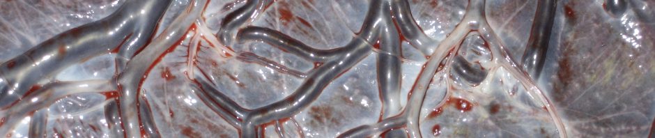
The placenta is compressed and the blood in the intervillious spaces (the interface between mother’s blood system and the placenta/baby’s blood system) is forced back into the spongy layer of the decidua (uterine lining). Retraction of the uterine muscle fibres constrict the blood vessels supplying the placenta, preventing blood from draining back through the maternal vascular tree (mother’s blood vessels feeding the placenta). This congestion results in the veins rupturing and the villi shearing off the uterine wall. A clot forms behind the placenta. The non-elastic placenta is unable to remain attached and peels away – usually starting from the middle.
At this point a small gush of blood may be seen as the placenta separates and the umbilical cord lengthens as the placenta moves downwards.
c)
After separation The placenta leaves the upper segment of the uterus and further strong contractions bring the walls of the uterus into opposition – compressing the blood vessels. At the same time the contracted uterine muscle fibres act as ‘living ligatures’ to the blood vessels running through them preventing further blood flow. An increase in the activity of the coagulation system means that clot formation in the torn blood vessels is maximised and the placental site is rapidly covered by a fibrin mesh.
As the placenta leaves the uterus the mother may feel the urge to push again and birth her placenta. Or, she may be far too busy with her new baby and the placenta will sit in her vagina until she moves.
This process is usually complete within an hour of the baby’s birth. However, sometimes it takes longer ie. hours… and hours. If you waited a long time to birth your placenta please post your story in the comments.
Here is an overview of how the placenta is born from my YouTube Channel:

Pathology – when it doesn’t work
The bottom line is that the birth of the placenta and haemostatsis (prevention of excessive bleeding) relies on effective uterine contraction. Ineffective uterine contraction is the main cause of post partum haemorrhage (PPH). The other causes are perineal/cervical damage, or even more rarely clotting disorders.
There are 2 main causes of ineffective uterine contraction after birth:
- Hormonal – Inadequate circulating oxytocin or inadequate uterine response to oxytocin. Inadequate response is often because the oxytocin receptors in the uterus have become saturated eg. by large doses of syntocinon over a long period of time during an induction (Belghiti et al. 2011; Phaneuf et al. 2000).
- Mechanical – something is in the way and the uterus cannot contract. Most often this is a full bladder taking up space in the pelvis and stopping the uterus from contracting down. It can also be a large clot in the uterus or a partially detached placenta.
Most PPHs occur after the placenta is out. PPH can and does occur after a c-section too – in fact it is more likely after c-section than vaginal birth.
Another complication can be a retained placenta ie. the placenta remains attached. The definition of a retained placenta varies – and I’m not game to put a timeframe on it. However, once you have done something (such as given an oxytocic drug – see below) you need to finish the job and get the placenta out. If you have not, and there is no bleeding or concerns about the woman, then… how long is a piece of string?
Active management of placental birth
In the 1950s syntocinon (pitocin) hit the birth scene. Syntocinon is an artificial version of oxytocin and is now used extensively for induction of labour, augmentation of labour and to ‘actively manage’ the birth of the placenta. It differs from endogenous oxytocin in the way it is released into the blood stream – ie. in a consistent dose rather than in pulse like waves. Syntocinon is also unable to cross the maternal blood-brain barrier and influence instinctive bonding behaviour.
When used to actively manage placental birth, syntocinon mimics the physiology described above by initiating uterine contractions. How active management is carried out varies considerably (Kearney, Reed, Kynn et al. 2019) and this drives midwifery students mad. Different practitioners do their own thing, and the literature is also inconsistent. Essentially syntocinon (10iu) is given to the mother by injection after the birth of the baby (although sometimes syntometrine). The cord is clamped and cut, and the placenta is usually pulled out using controlled cord traction. The order and timing of these interventions varies, although obviously pulling the placenta out comes last. The areas of debate/negotiation are:
- Timing of injecting syntocinon: Originally syntocinon was given with the birth of the baby’s anterior shoulder. Nowadays, it seems to be given after the birth of the baby. There is no research determining the best time. Syntocinon takes around 3 mins to work when given IM (into muscle) – so in theory to mimic physiology it probably should be given soon after the baby arrives. However, there is no evidence to support early administration of syntocinon. In fact, the research suggests that giving the oxytocic before or after the birth of the placenta makes no difference to the risk of PPH (Soltani et al. 2010 – Cochrane Review).
- Timing of clamping and cutting the cord: The risks of premature cord clamping are now well known, and a Cochrane review recommends delaying cord clamping. Most midwives I know (regardless of where they work) wait until the cord has stopped pulsing before clamping. Some are concerned about syntocinon crossing the placenta into the baby. However, syntocinon does not cross the placenta as previously thought. There is also a theory that the strong contraction caused by syntocinon will shunt excess blood from the placenta to baby. Again there is no evidence that this happens and during physiological birth all of the blood transfers to the baby – there is no ‘excess’ blood to shunt. There is also no evidence that waiting to clamp the cord increases the risk of jaundice (Rana et al. 2019)
- Whether to ‘drain’ the placenta: If the cord has been prematurely clamped, some of the baby’s blood is trapped in the placenta – this makes the placenta bigger and more bulky, and in theory/experience more difficult to get out. There is no research to support this… but many midwives will leave the placenta end of the cord unclamped and drain the trapped blood prior to attempting to deliver the placenta. Personally, this is my preference as I notice it is much easier to get an empty placenta out. Something I learned while collecting cord blood. Of course, it is even better if all that blood is in the baby.
- Whether or not controlled cord traction (CCT) is used and when: It is standard practice to pull the placenta out after syntocinon has been injected, and the umbilical cord has been cut. Some midwives wait until they have seen signs of placental separation before pulling (trickle of blood and lengthening of the cord). I think this part of active management causes the most problems. If you pull on a placenta that has not yet separated you can partially detach it = some blood vessels are ‘torn and open’ but the uterus cannot contract because the placenta is in the way. Or, you can detach it before the syntocinon is working i.e. no contractions to stop the bleeding. Or worse case, and very rare scenario you can pull the uterus out (inverted uterus)! You can also, more commonly, snap the umbilical cord – which often freaks everyone out. But a snapped cord is not a big drama. It just means the mother will have to get up and push her placenta out… Which brings me around to the idea of not pulling at all. A study by Gülmezoglu et al. (2012) found that the ‘omission of controlled cord traction’ did not increase the risk of severe haemorrhage (they only looked at severe). And another study found that CCT made no difference to the PPH rate and concluded (Deneux-Tharaux et al. 2013). So, women should have the option of getting upright and pushing, or having someone pull their placenta out for them. Or even perhaps pulling their own placenta out?
Active management is usually (not always) quicker than physiological. This is probably another reason it is favoured in hospital settings. Less time waiting for a placenta = less time stressing out about a potential PPH, and you can get the woman to the next station (postnatal ward) quicker.
Occasionally syntometrine is used for active management. This is a mix of syntocinon and ergometrine. It is not generally used nowadays because the ergometrine acts on smooth muscle – all smooth muscle. Therefore the side effects are vomiting, raised blood pressure and potentially a retained placenta due to the cervix shutting… although I’m not convinced about the cervix closing firmly enough to trap a squishy placenta.
What the research tells us – and doesn’t tell us
The physiological vs active management of the ‘third stage’ has been going on since I was a student midwife (I did a literature review on it as an assessment). Today I am doing it the easy way and relying Cochrane to review the studies for me (Begley, et al. 2019; Salati et al. 2019). In summary, the reviews note that there is a ‘lack of high quality evidence’ but conclude that active management reduces the risk of haemorrhage in a ‘mixed risk’ population birthing in hospital. They also raise concerns about side effects – increased blood pressure, afterpains and vomiting (probably due to the use of syntometrine in some studies); reduced birthweight for baby (probably due to reduced blood volume following premature clamping); more women returning to hospital with bleeding (?). In regard to the last side effect – anecdotally, midwives report greater blood loss on the postnatal ward after the syntocinon or syntometrine has worn off but this is not measured in studies.
Both Cochrane reviews state that active management is not effective at reducing significant PPH for low risk women. When interpreting the review findings it is important to remember that all of the studies included were conducted in a hospital setting. The experimental group were those having ‘expectant’ management, and the practitioners attending the ‘physiological’ placental births were most likely doing something that was not their usual practice, and they may have been unprepared for, or uncomfortable with this approach. Care providers in hospital settings can be inexperienced at supporting physiological placental birth (Reed, Kearney & Gabriel 2019; Kearney, Reed, Kynn et al. 2019).
Women with care providers experienced in supporting physiology have very different outcomes. A study by (Fahy, et al. 2010) compared active vs holistic physiological care. The midwives in the study were experienced with physiological placental births. In contrast to previous studies, active management was associated with a seven to eight fold increase in PPH rates compared to a holistic physiological approach. Another retrospective study (Davis et al. 2012) found a twofold increase in large PPHs (1000mls+) for low-risk women having an actively managed placental birth in New Zealand compared to those having a physiological placental birth. In summary – for women having undisturbed physiological births active management of the placenta increases their chance of having a PPH.
As previously described, the baby plays an important role in assisting with the birth of the placenta. Therefore, undisturbed interactions between mother and baby are important in avoiding a PPH. A recent study looked at the impact of removing babies from their mother after birth and found that: “women who did not have skin to skin and breast feeding were almost twice as likely to have a PPH compared to women…” who did have this contact with their baby (Saxton et al. 2015). The authors conclude: “…this study suggests that skin to skin contact and breastfeeding immediately after birth may be effective in reducing PPH rates for women at any level of risk of PPH.”
Back to my initial title statement
A safe and effective physiological placental birth requires effective endogenous oxytocin release. This is facilitated by:
- A physiological birth of the baby: No interventions during the birth process eg. induction, augmentation, epidural, medication, instructions or complications.
- An environment that supports oxytocin release: Privacy, low lighting, warmth and comfort. No strangers entering the birth space eg. paed or extra midwife.
- Undisturbed skin-to-skin contact between mother and baby: others must not handle the baby or engage the mother in conversation. These mother-baby interactions may result in breastfeeding, but this should not be ‘pushed’ as not all babies want to breastfeed immediately.
- No fiddling: No feeling the fundus (uterus). No clamping, cutting or pulling on the umbilical cord. No clinical observations or ‘busying’ around the room.
- No stress and fear: Those in the room must be relaxed. The midwife needs to be comfortable with waiting and have patience. The mother must not be stressed as adrenaline inhibits oxytocin release. This is why a PPH often occurs after a complicated birth (eg. shoulder dystocia) and when the baby needs resuscitating.
- No prescribed timeframes: Many hospital policies require intervention within half an hour if the placenta has not birthed. This is not helpful and generates anxiety which is counter productive.
The most important factor in ensuring a safe physiological birth of the placenta is a physiological birth of the baby.
However, in Australia (AIHW 2021) less than a quarter of women go into spontaneous labour and continue to labour without augmentation. Out of that % how many labour without an epidural or other medication? Out of that % how many are birthing in the conditions described above? I pose the questions because these stats are not presented.
An interesting study by Nove et al. (2012) compared PPH rates between planned hospital birth vs planned homebirth. They adjusted for co-founders such as risk factors associated with PPH. The study found lower rates of PPH for women planning homebirth, even if transferred to hospital during labour or afterwards. The authors conclude: “Women and their partners should be advised that the risk of PPH is higher among births planned to take place in hospital compared to births planned to take place at home, but that further research is needed to understand (a) whether the same pattern applies to the more life-threatening categories of PPH, and (b) why hospital birth is associated with increased odds of PPH. If it is due to the way in which labour is managed in hospital, changes should be made to practices which compromise the safety of labouring women.”
Summary
Active management of the placenta will reduce the chance of a PPH in a setting that does not support physiology and in which routine intervention is the norm. There are further options within active management that can be negotiated (see above). Physiological placental birth is an option, and possible if you manage to avoid induction, augmentation, an epidural or complications – but be aware of how difficult it may be, and don’t beat yourself up if it doesn’t happen.
Further reading/resources
- Birthing the placenta: women’s decisions and experiences
- Placentas and cord blood – The Midwives’ Cauldron Podcast
- Can I have a natural placental birth after induction? – Sara Wickham
- Hastie C, Fahy KM. Optimising psychophysiology in third stage of labour: Theory applied to practice, Women Birth (2009), doi: 10.1016/j.wombi.2009.02.004
- Placental birth: a history – PhD thesis Stojanovic 2012
- 30 Minute Third Stage – Gloria Lemay
If you enjoyed this post, you can find more of my work in the following resources:
Join my Mailing List to stay up to date with new content and news
Learn more about Childbirth Physiology in my Online Course


Hi, I’m a first year student midwife and I actually just finished an essay on this exact topic yesterday.It’s interesting when reading the studies, the limited knowledge and experience some midwives have these days of true holistic physiological birthing of a womans placenta if they have been in a hospital setting and lost or never learnt the proper process. I suppose that is just another example of the medical-model of care seeping in through the birthing suite doors where holistic midwifery care should be practiced on low risk women in normal labour. Thanks for the interesting read 🙂
My first was actively managed (I didn’t know the difference then), but with my second (at NGH) I opted for physiological. After a natural (very quick) birth, I had just husband, Midwife and student in the room. baby suckling well. After his birth I had no contractions at all. We waited one hour and then I had the injection and some massage, placenta was out in 2 contractions. At first I was dissapointed that it hadn’t happened the way I wanted but each birth has been a learning curve for me and now I now more about how this last part happens! We’ll see how #3 goes!
In my experience at homebirths 1 hour+ is normal. This is the problem with hospital timeframes. Very few women manage to meet the arbitrary limits made up by hospitals. It is shame that you were unable to be supported and trusted.
My first birth was in a hospital with a severely hyperactive doctor (waters broke, 24 hours of no contractions, then 7 hours of irregular, 5.5 hours of regular, and 1.5 hours or pushing contractions followed by vacuum – my midwife was extremely irritated). He wanted the placenta out within 20 minutes and insisted on having it actively managed (controlled cord traction, no injection). It came out fine, thank goodness.
My second birth was a water birth at home (onset of regular contractions followed quickly by waters breaking, 3.5 hours of regular contractions, 15 minutes of pushing contractions) with the same midwife. I got out of in the tub for about an hour after the birth (it didn’t seem that long to me, but I’m looking at the midwife report now). At that point my midwife’s notes say “placenta appears to be separated”. I pushed the placenta out about 90 minutes after the birth, standing over the toilet (the midwife caught it). No complications. The cord was only cut after the placenta was out. Either my midwife was completely unconcerned about the length of time or she has a fantastic poker face (actually, I know she has a fantastic poker face, but she was probably also unconcerned).
At the birth only my midwife and husband were in the room. We were joined a few minutes later by my elder son (2 years old) and my sister. I don’t know if that made any difference in the time to birthing of the placenta. I do know I wanted them there!
I am much influenced by Kathleen Fahy’s (et al) work in Women and Birth (2010) on “Holistic physiological care compared with active management of the third stage of labour for women at low risk of postpartum haemorrhage: A cohort study”
Kathleen Fahy *, Carolyn Hastie, Andrew Bisits, Christine Marsh,
Lurena Smith, Anne Saxton
It is a great study isn’t it… I have discussed it in the post 🙂
Interesting, I definitely feel the atmosphere in the room is paramount, and often send women out to the bathroom with a Pan, Women need privacy for their placenta.
toilets are a great place to birth babies and placentas 🙂
I looked after one woman who always went into the shower by herself to birth her placentas. Came out of the bathroom with the placenta in a bowl
For my son’s birth in January, I requested a physiological 3rd stage. The hospital Obs wasn’t keen but my midwife on the day had no problem. My labour was spontaneous and intervention free and I laboured and birthed standing up. When the time came to birth my placenta, the midwife got me up on the bed while I tried to attach my son to my breast. I contracted a few times but she was gently pulling on the cord. The placenta was birthed about 40 mins after my son. Up on the ward, I was haemorrhaging. A syntocinin drip was started and I was told to fast for an EUA. After feeding my son again I went to the toilet and passed a large mass of membranes.
… one midwife told me that the PPH was my body trying to expel the membranes which made sense to me as it stopped after that happened. But all the other midwives were telling me it was because I refused the synto after delivery. “Oh, you’re the one causing everyone to panic.” I’m just curious to see what your opinion is. My labour was a 6.5 hour VBAC and my first time in labour. No interventions, and a completely hands off, drug free birth, complication free preg, 1st C/S for breech. I
Hi Violet,
I hope you don’t mind me replying to your question, but your experience is the perfect example of a midwife who wasn’t familiar with holistic physiological placental birth. She shouldn’t have been pulling on your cord, not even gently, not even a little bit. That *may* have been the cause of your PPH.
She absolutely shouldn’t have been pulling on the cord. But I don’t agree that this is likely the cause of your haemorrhage. It is common practice in the US to use CCT and give an ecbolic after the placenta is expelled. Their stats for haemorrhage don’t differ markedly from ours. However a midwife who is unfamiliar with physiological management may not be familiar with the separation bleed that accompanies a physiologic third stage and may have panicked a bit. I often suggest that the woman puts traction on the cord if it seems that traction might be necessary, on the basis that a woman with a placenta still attached would most likely not pull if it hurts.
I have to agree with Holly. Pulling on a cord is not part of a physiological approach to birth. There is potential to interfere with the separation of the placenta and tear the membranes = bits of membrane are left behind and interfere with contraction = bleeding. However, is certain circumstances if you are ABSOLUTELY SURE the placenta is separated and sitting in the lower part of the vagina, and the woman is unable to push it out, the woman can pull, or you can pull gently. NEVER PULL ON AN ATTACHED PLACENTA during a physiological process.
this is a case of using spittle to bathe in the presence of ocean. it is equivalent to taking unnecessary particularly in a woman who underwentVBAC
I’m not so sure with the link you make between SD and PPH. My understanding is that it is less to do with stress hormones (although I’m sure they play a part) and more to do with a tired uterus which has been contracting without effect to expell the baby, and an often larger placental site. Also have you seen the article published in this journal, which basically says that physiological management doesn’t increase severe haemorrhage.
BTW, four t’s….tone, tissue, trauma, thrombin.
There is a link to SD and PPH for a number of reasons… eg. the placental site may be bigger as the baby may be bigger, a ‘tired uterus’ is a theory too (although I don’t buy it as often SD occurs after a quick labour). Not sure which article you are referring to? I have used Fahy et al and included the findings.
I don’t teach the four T’s because I find students get confused by it. Essentially the vast majority (80%) of PPH cases are due to ‘tone’ – tone is effected by ’tissue’ i.e.. the placenta but the issue is still a ‘tone’problem. It is the tissue preventing the tone. I was taught the 4 T’s but don’t really find it helpful in the moment or for teaching.
http://www.midwife.org.nz/index.cfm/3,114,279/nzcom-journal-41-lo-.pdf
Sorry forgot to post the link.
Have cared for several woman where the uterus is well contracted, so tone not an issue, but clot caught in the cervix is, I definitely find the 4ts useful when thinking about a haemorrhage.
Thanks for the link. I haven’t found a clot in the cervix to be a problem re. Bleeding if the uterus is contracted and I’m not sure what the mechanism would be. Where’s the blood coming from if the uterus is effectively contracted? Women can feel pretty awful when a clot or the placenta is sat in the cervix – not sure why. Interesting. I’m pleased the 4 Ts work for you. They don’t for me. 🙂
I had a large retroplacental clot sat in my cervix following the birth of my second baby at home which caused me to go into cervical shock.
I’ve had 3 babies, all born at home. All 3 times my placentas took a while to be born, although I suspect they had detached completely and were just sitting in the vagina for a while.
Baby #1: placenta took 2 1/2 hours to be born. I was having the world’s strongest afterpains the whole time and finally I was ready to get the placenta out. I sat on the toilet and gave the cord a good tug because at that point I was pretty sure the placenta was detached, but just stuck in the vagina. Estimated total blood loss = 100 cc.
Baby #2: After about an hour of totally uninterrupted skin-to-skin in my bed (in a nice, cosy warm room with a midwife who peeked in to check on us but didn’t disturb me at all), I was ready to birth the placenta and take a shower. The midwife checked to be sure it had detached (yep) and gave a small amount of traction while I gave a little push. Very minimal blood loss again.
Baby #3: about the same scenario as last time. I snuggled and nursed the baby for about an hour, then I wanted to get the placenta out so I could shower. Again, I gave a push–always harder to get the placenta out than I think it should be!–and the midwife did very gentle traction after determining the placenta was detached. Estimated total blood loss = 15cc. yes, that’s right. She was amazed and took a photo so she could prove how little blood there was!
I agree that it’s a very different matter to have a “physiological” 3rd stage in a hospital compared to in a birth center or home setting, and I suspect that’s why the Fahy study had such different results. It’s not just about delayed cord clamping and waiting for the placenta and not giving a routine shot of Pitocin; it’s about uninterrupted skin-to-skin, having a warm room and minimal level of distractions, etc.
Thanks for sharing your experiences with physiological placental births 🙂
Thanks for commenting that to you the placenta seems harder to get out than you feel it should. I felt that birthing the placenta was more uncomfortable and the pushing more painful than the birth, where I felt pushing didn’t hurt at all. I had been lead to believe that the placenta basically births itself and just flops out – so NOT true in my case!
I agree – I had no idea how much the birth of the placenta would be a real contraction and a real push. (I also was really shocked about how much it hurt to birth my babies shoulders, I didn’t feel that was at all fair – I’d already got her head out).
What are your feelings about delaying cord clamping around 10-20 minutes (waiting for a white cord) with modified active management?
Where mum is continually observed for pv loss. Surely that’s a better option?
In my experience cutting bulging pulsating cords causes pph.
Continually observing pv loss how is that giving the woman privacy.
It can be done discreetly !
Have you read the book Promoting Normal Birth? There is some very interesting research in there on a natural physiological third stage. Let me know if you want a copy of the study.
Amy V. Haas, BCCE
I posted one here: http://www.facebook.com/note.php?note_id=292276417510166
The book has a more complete analysis, is fairly recent, and agrees with your theory.
This is a link to purchase: http://www.amazon.com/Promoting-Normal-Birth-Reflections-Guidelines/dp/1906619255
And lastly, This article is older: http://www.facebook.com/note.php?note_id=288622841208857
but well done.
Thanks for the links. I have read this book and I recommend it to anyone involved with birth 🙂
Thanks, great reading .
As a doula, I have supported women who have chosen to wait, and so far, 2 hours has been the longest, a few homoeopathic remedies and a walk to the toilet ususally help. But how to get the privacy, peace and quiet in a hospital??
I waited 3 hours for my placenta at my first home birth… I think a combination of full bladder, too much noise and being cold didn’t help, but I still bled a lot less during the birth and afterwards, and with much less lochia, than the births with synto…
It took about 3 hours for my placenta to detach with Homebirth baby number 3. One and two were actively managed but natural births. After an hour flew by and the placenta had not detached I spent some private time on the toilet after taking some homeopathic remedies. I walked around a bit, had a shower, tried a gentle tug myself….nothing. I had nursed the baby several times, visited with my older children then went back to the toilet. Out came the placenta (onto a chux pad that created a bowl) and finally was able to pass urine. Perhaps the full bladder was the problem? don’t really know. My midwife was very calm and I barely lost any blood.
The full bladder is the usual culprit. However, breastfeeding your baby and visiting your older children probably released a whole load of oxytocin.
I’ve had a managed third stage with all 3 of my labours. My first 2 were inductions, the last was a natural waterbirth. With my 1st it was a syntocinon induction and they assumed that as I still had the drip up it would expel the placenta, it didn’t and they gave me syntometrine. I lost around 300ml, so have been advised to have a managed 3rd stage for each subsequent delivery. Interesting to read that I may have been better not doing with my last one! With my last one I had 2 shots of syntocinon and the placenta still wasn’t fully detached and boy was I bleeding. I finally got my little one latched on and that did the trick!
My daughter was born at home at 37 weeks. She came out a little shocky and surprised, but was in very good health. My placenta took another 2 and a half hours to be ready to come out. I believe that it just needed a little extra time to release and let go, considering that Sweet Ayla came so early. I am so grateful that I was comfortably at home with a loving and patient Midwife and that no intervention was needed to rush out the placenta.
Pingback: L’expulsion du placenta: plein d’infos de The thinking midwife « Annie l'accompagnante
Thank for this great blog really enjoyed reading it – I agree.
Wow, of all of the birthing processes, I have taken the placenta birth for granted the most. I’ve had three home births, and every time the placenta came out easily, on it’s own, and shortly after the birth. Each time I left the birthing tub with my baby, sat on a birthing stool to nurse, and, plop! There it is! This post gave me something to think about. I’ve been spoiled.
Hello delicious women… I am a Menstrual Educator, I share with women the cultural tools to bleed well and strong, and from there, birth well and strong… I have a physiological question about the the uterine wall as it releases the placenta… are there any studies about the cellular level of change in comparison to menstrual release and cessation? Is the body doing essentially the same thing is does every cycle? Bleed and then stop bleeding when it is time? Personally I find the placenta an incredible organ that needs a lot more understanding than we have given time to. Thank you for a really well written blog, I am sure I will enjoy reading more!
I experienced a serious PPH after a partial separation of my son’s placenta..& it was retained…quite adhered to the uterine wall…..the thinking is that when I moved quickly between the time of birth & then trying to birth the placenta..I lost the retro placental clot & thus there just wasn’t enough behind that placenta to bring it down & out…possibly a few factors involved but it was interesting considering it was a beautiful, unhindered, unmanaged home birth which went a little haywire…xx
Sometimes things do go a little haywire regardless of the situation… it’s the unpredictable nature of birthing and of parenting 🙂
I am shocked that only 21% of women go into labour spontaneously. If that is so it is a very serious situation that needs to be addressed in its own right. Women have to say no to such a ridiculous situation. It certainly will have an impact on third stage.
Let’s get a few parameters sorted.
The placenta is the baby’s. It is not the woman’s. It is an organ from the same beginnings as the rest of the baby’s body.
There is no such thing as ‘delayed’ cord clamping. All cord clamping is precipitous. The only reason that cords were ever clamped was to protect hospital linen from the precious cord blood that was meant for the baby’s body soiling the sheets because the cords were cut before they had stopped delivering the blood to the baby. Considering that the blood in the cord is 30-50% of the baby’s total blood supply it could make quite a mess. It was about laundry costs =$$$. When a cord has stopped pulsating there is no need for clamps. It can simply be cut with very little blood present. Clamping a cord that is still pumping causes a great disruption to the baby and mother’s physiological situation. There was no research or controlled trials conducted before this protocol was introduced.
Most of us are living with profound cord and placenta trauma. It has become so assimilated into how we experience ourselves that it’s damaging fallout has come to be regarded as ‘normal and we have developed other accounts to explain difficulties that are actually originating from our cord trauma’. Most ‘information’ written about ‘third stage’ is redundant. The discussion is about something that should not ever occur. ‘Third stage’ like ‘braxton hicks’ and ‘birth canals’ are male constructs about a female experience that objectives and confuses the issues. Having said all that, we must consider the compromise in the physiology of both mother and baby when pharmacutical drugs are part of the equation.
To appreciate the forces present at birth we must begin at the beginning. This is the moment of implantation 9mths before. At implantation two crucial things occur simultaneously. The mother accepts the pregnancy and the fertilized ova commits to incarnation. This is a most useful understanding about the mother child relationship. It takes two to tango!
The environment at this time provides a most powerful cellular imprint that will always reverberate throughout this most primal relationship of mother and child. These elements occur across the physical, mental, emotional, energetic and spiritual planes of existence and all need to be considered.
Our relationship with our placenta is profound. The placenta itself is an organ of high intelligence. The second edition of my book ‘Lotus Birth’ has an additional 80 pages that covers some very exciting understandings about our placenta and the long term effects of how we experienced our time with it and how we can heal. Our limbic systems are still coping with that early trauma. Get your copy http://www.lotusbirth.net
Third stage provides a unique time.
I have seen mothers hold on to the placenta when they have needed more time. Sometimes there can be ambivalence about no longer being pregnant. If it has been a very fast birth she may need time to regroup before moving to the next stage. If it has been an unexpected pregnancy, particularly very close to another child she may create a gap before moving on. Sometimes it may be connected to her relationship with the father. It may be related to her own birth. It may be as far back as her own conception.There are many scenarios that can present and if understood and allowed to be revealed and resolved then placentas suddenly plop out.
A midwife friend told me of a mother who had kept the placenta until the next day (the cord had been cut)and while standing at the kitchen sink let it go
I was once called to attended a birth where the placenta was still inside the mother 5hrs after the baby had been born. The baby was fine. The mother was fine but the placenta was still inside her. It was a lotus birth so the baby was still intact with its cord. Many strategies had been tried but the situation had not changed. As I sat with the mother and asked her what her prevailing thought was she said that she kept thinking about Siamese twins. So we explored the possible advantages and disadvantages of being a Siamese twin. As you can imagine there was much about to-ing and fro-ing, attachment and co-dependence, always having somebody there, never having time to yourself, and other related phenomena. We spoke for about 5-10 minutes and then the placenta simply came out. Of course it probably had nothing to do with actually being a Siamese twin, although I’d not discount it entirely. Her unconscious had provided her with a very effective metaphor that had enabled deep and complex issues to be resolved. Metaphor is the language of the unconscious and it is our deep unconscious forces that determine what occurs .
.So Active management becomes ‘necessary’ when there are unresolved issues which manifest physiologically. This highlights the great benefit of the midwifery model of continuity of care where during the healing environment which is pregnancy many issues are given space to present and are resolved before the birth time.
Your conclusions are echoed by ours Rachel & thanks for mentioning our study. When women have intervention in labour, then active management is warranted. However, if a woman births her baby in a straightforward way, then providing the right environment for her to birth her placenta under her own steam means she is much more likely to have minimal blood loss. I’m not sure if you have seen our theory into practice paper, so I include it here for those who are interested.
http://scu-au.academia.edu/CarolynHastie/Papers/110837/Optimising_psychophysiology_in_third_stage_of_labour_Theory_applied_to_practice
I’m fascinated by the placenta and the way that women give birth to that fabulous organ. I love that photo you have on this blog post of the woman birthing the placenta into her own hands.
I wrote a blog post about how the placenta is so often missing in birth films as it irks me that women don’t often know about the importance of that part of labour and that they are still in labour until the placenta is born http://thinkbirth.blogspot.com.au/2010/05/wheres-placenta-in-birth-films.html
One notable exception is the brilliant 1979 Birth in the Squatting Position film from Brazil http://www.youtube.com/watch?v=gI6q0nB8VgM&list=FLNxfEMKGu_bfnGQKoRSqkPg&index=1&feature=plpp_video – this video changed my midwifery life when I first saw it in the very early 80’s. There is an excellent placental birth in this film.
One of my most interesting experiences about third ‘stage’ was in Papua New Guinea last year. I was working with students in a labour ward that saw a full classroom (40+ children in PNG – big classes there) of children born every day. When we arrived at work early one Monday morning, I discovered there were no artificial oxytocics to be found anywhere – they had run out the previous afternoon. I noted there were NO incidences of excessive blood loss recorded in the birth register overnight. Now you have to know that this is a country where maternal health is very poor and the death rate is very high, most often caused by postpartum haemorrhage. I asked one of the night duty midwives what did they do overnight with no oxytocics – the midwife answered with a nonchalant shrug of her shoulders, “we just put them on the mother’s chest (skin to skin) and let them breastfeed” – no, that is not standard practice there and yet they knew what power there was in doing it. If ever the amazing properties of the skin to skin experience of mother/baby and unimpeded breastfeeding was evidenced, surely that was it!
Thanks so much for your comments and links. The film is amazing… you are so right about the invisible placenta in most birth scenes.
I have added the ‘theory into practice’ paper to the blog post. 🙂
I have been present at a hospital birth when there has been 7 hours before the placenta was released. The dad was very insistent the doc had hands off and as mum wasnt compromised at all we waited and waited not sure this would happen now but it was another part of learning from women
Thanks for your write up. I really liked your description of what it takes to have a physiological birth. I’m a Lamaze childbirth educator in the US, and had been wondering, too, how most effectively to share info about the third stage in my classes. I learned about the 4 Ts, but I prefer to primarily talk about the main contributors to third stage bleeding and divide them into two categories — one of factors that the mom might have some control over and the other category of known contributors that the mom generally has less or no influence on. And it’s helpful to let moms know that management of the third stage is kind of like an “if . . . then” statement — if the first stages of labor are physiological, then the third can be physiological. If the first stages of labor are more medically managed, then the third stage is perhaps better managed medically, too. This gives the moms who want a physiological birth even more reasons to prepare for physiological birth with caregivers who are experienced in this. Here’s a link to the 2-part write up I did for Lamaze on it. http://www.scienceandsensibility.org/?p=4103
Thanks – I’ve added your write up to the blog. I like the ‘if… then’ concept 🙂
Thank you for another excellent posting. Can I ask you to expand a little on this “…there is a theory that the strong contraction will shunt excess blood from the placenta to baby… I’m not so sure. ” please? That was my assumption also.
Expansion…
There are a few questions raised by this assumption.
1. we don’t know whether the syntocinon creates a stronger contraction than oxytocin. Endogenous oxytocin creates a pretty strong contraction to facilitate birth of the placenta.
2. The physiology of a big contraction shunting blood is questionable. In labour large contractions interrupt blood flow, not increase it.
3. Whilst the cord remains intact distribution of blood back and forwards continues until the cord stops pulsing. There is even a theory that the cord vessels actually regulate this blood flow (in pregnancy and labour). So, perhaps an initial ‘shunt’ of blood would be sorted out by the placenta/baby circulation.
4. There is only so much blood in the baby/placenta unit. You cannot make a bigger volume. Physiologically the vast majority of this blood would end up in the baby: http://midwifethinking.com/2010/08/26/the-placenta-essential-resuscitation-equipment/… so a ‘shunt’ of blood may not make a different overall and may be beneficial if followed by immediate cord clamping.
5. Syntocinon takes around 3 mins to work by which time the cord is usually slowing or stopping blood flow, especially if it is not given immediately which it often isn’t.
So… that’s me thinking aloud. I think a lot of the things we assume to be true about birth are the things we are told and take for granted. Once we start thinking and challenging ourselves about what we know it all becomes far less clear 🙂
Once you have witnessed a baby with its placenta intact until it comes away without interference and have seen the natural order with your own eyes much of the discussion becomes obsolete. Robin Lim has some wonderful utube offerings. She tells of resuscitating a baby with its cord still intact by massaging the placenta in warm water. There needs to be a revolution in how people act at this time. Babies need their placentas and mothers are best served by their babies’ needs being met. I think that the problems that have become associated with third stage are actually caused by the inappropriate protocols that are in place. They are part of the cascade of interventions. It must be awful to have to work in such an environment.
I have witnessed what you are describing. I attend homebirths and the placenta remains attached to the baby until the mother decides (or not) to cut the cord – usually well after the birth of the placenta.
Because most women are birthing in hospital environments I believe it is important for them to understand what this means in terms of how it alters their birth experience. I end up debriefing too many women who thought that they could enter the hospital and have a physiological birth, failed and then blamed themselves rather than the environment. Also, most midwives also work in these environments and the discussion and debate must carry on if we want change.
Agreed Rachel. It is a good and most necessary conversation.
My first was born in a hospital (super active management w/pre-e) and there were so many things I wish had been done differently. Nothing I asked for ahead of time happened, like delayed cord clamping, getting to hold the baby and breast feed right away, and no tugging on the cord to pull out the placenta. Luckily I didn’t have PPH, but they likely gave me something to prevent that. My second, I had at home, just me and my husband. I had read (I think in “Pushed”) that in Jamaica (again, not sure if I’m remembering the country correctly), when care providers were taught to do immediate cord clamping, the rates of PPH skyrocketed. We decided to wait and cut the cord more than an hour after the birth. It felt incredibly peaceful and right. Of course, most people get a little weird when I tell them my husband used the kitchen shears 🙂
In many traditional cultures the umbilical cord is cut by a tool associated with the father’s trade eg. a spear or scythe 🙂
Should I let my partner cut our baby’s cord with his chainsaw then? 😉
I had a homebirth, it was maybe 45 minutes before the midwife was starting to actively manage the placenta, it was still firmly attached so she gave me the shot, did some tugging and I guess I was bleeding from the tears, so she gave me another shot and told me that if it didn’t come out we may need to transfer because of blood loss, finally after one big push from me it came right out. I hope this next one will be much less dramatic and natural. I will be giving birth in a birth center where the midwife is very hands off but their policy says 30 minutes. I hope my body can do it in that amount of time!
You have to give consent for any intervention. If you get to 30mins you can decline active management. It might get a little stress and the worst thing for you at this time is confrontation… but you do not have to have anything you don’t want.
I had a homebirth with my second child and the placenta took ages to come out – 2/3 hours+ and even then I got no real urge to expel it. It seemed very reluctant to come out when it did appear which turned out to be because I retained a lot of it (removed under a spinal a few hours later in hospital). I’ve yet to have my afterbirth debrief with the hospital but I’m really interested/curious as to why it didn’t come away naturally all and how this will affect any future pregnancies.
Just because this happened once does not mean it will happen again. I have been involved with lots of successful placental births following a previous retained placenta 🙂
Good to know :). Can you tell me are there any possible problems from the manual removal itself? In all the euphoria of the birth and just wanting it done and to get home again I forgot to ask the doc!
Only during the actual procedure or short term ie. damage to the vagina/cervix; infection; pain etc. Not long term 🙂
Manual removal of the placenta is associated with Asherman’s syndrome (and especially so if following the manual removal they do curettage “blind” – that is, without ultrasound guidance – to ensure they’ve got it all.)
My first child was born via c-section due to pre-ecclampsia. I had a HBAC for my second child. We waited about 6 hours for the placenta to be delivered. After a few hours, the midwife tried an experimental shot of pitocin into the cord (which had been cut when it had stopped pulsing). About an hour later, we tried a shot of pitocin in the thigh, which also didn’t work. Gentle traction yielded pain. So we went to hospital. The hospital staff told me I was still 10cm dilated (which didn’t surprise me, labor hadn’t finished, right?), and my blood test showed signs of early infection, which they attributed to the dead placenta. They performed a manual removal of the placenta, which thankfully, was only lightly adhered to the uterus… probably at the site of the prior c-section scar. Best case placenta accreta, I suppose. They then performed a “light curettage” to ensure they hadn’t missed anything.
That procedure is the most likely cause of the Asherman’s syndrome I now have, diagnosed after physical investigation following two early miscarriages in row (one at 6 weeks gestation, another at 9 days gestation) following the birth of my second child. I also experienced tail end brown bleeding following menses up to day 11 of the cycle, when cycles resumed after the birth of my second child, which was a clue along with the miscarriages. I’m now looking at surgery to repair the uterus, and hope it works.
Not to be all doom and gloom, but I just wanted to make it known that there are some long-term risks associated with the manual removal of the placenta.
Thanks for sharing this important information. I hope your surgery is successful.
My story of prolonged physiological third stage, since you asked – had my second baby in a birthing centre, but no interventions and no medication, all quick and easy. Placental delivery took three hours. (Longer than my time in active labour – never know quite how to answer the question about how long my labour was!) I did eventually have the Synto and the placenta came out a while after that when it kicked in. Ironically, one of my reasons for refusing the Synto initially was because I wanted to avoid possible side-effects – I wish I’d realised that the side-effects of a prolonged third stage are also pretty darned unpleasant. I had horrible cramps and nausea. Oh, well – came out all right in the end anyway, but if I’d known then what I know now I’d have had the Synto as soon as the cord was clipped. (And, in another irony, the midwife cut the cord immediately after birth before I’d had a chance to tell her not to, so I lost the other advantage I’d been hoping to gain from physiological third stage, of being able to delay cord clamping.)
A postcript to this: When I posted my birth story on the group I was a member of, one woman told me she’d heard that upright position immediately after birth helped avoid this sort of problem. I don’t know whether this is true or not, but do know that I had a straightforward physiological third stage with my first birth, and that I probably was in a more upright position straight after the birth with that one (not deliberately so, but I was on all fours for 1st delivery and in left lateral for 2nd delivery, so I think I would automatically have ended up in a more upright position when I was turned over the first time after giving birth). Maybe a coincidence, but maybe not – I do know that if I was planning to have a third baby, I would make sure I was in a reasonably upright position after delivery!
Gravity might help. Having said that – I don’t think women should be hassled into positions after the birth of the baby. It is more important that they relax and spend time with their baby as this will assist with separation of the placenta more than gravity.
I just had my first baby at home 7 weeks ago! My placenta took about an hour and a half to come all the way out. When it did start coming out, it came maternal-side first, and only about 3/4ths the way out. my (AWESOME) midwife held it while I knelt so it wasn’t pulling on me, and slowly twisted it until it came all the way out. She said if I felt any pain or pinching to let her know immediately and she would stop. The midwives were interested because it came out maternal side first, and still seemed to be attached to something. Not once did they even touch my fundus or mention anything about using herbs or anything else to help it along. They were relaxed and not worried, so neither was I. It was a great experience.
That type of placental birth is often referred to as a ‘dirty Duncan’. It happens when the placental separates from the edge first rather than the middle. If you look at the diagrams you can see that when the placenta separates from the middle it comes out membrane side first. If it slides out sideways the first thing you see is the maternal side and it can be trickier to birth as it is not quite a ‘neat’. 🙂
Many years ago on the ICAN list, there was a woman who had a UC and waited over 40 hours for her placenta. She wasn’t bleeding excessively, so she just waited. If I remember, she believed that she’d had a bit of an accreta and that the extra time allowed the placenta to detach on it’s own. Not sure if that’s even possible, but I though it was interesting.
As for me, first baby was a cesarean. I hemorrhaged due to excessive pitocin prior to surgery; my uterus did not want to contact any more after the surgery. No one said anything to me, of course, but from the bits of conversation I heard, things were a bit tense. 2nd baby was a UC. Waited for quite awhile after birth to cut the cord (probably at least an hour) and placenta still wasn’t out. Cut the cord, took a shower, had supper (tried feeding the baby a few times in there). Eventually decided I needed to go to the bathroom & it just fell out in the toilet. I have no idea how long it had just been sitting there. 3rd baby was a water birth, started getting contractions within half an hour and pushed it out. 4th baby it was taking awhile again & I really wanted to get out of the pool, so I tried very, very gently pulling on the cord to see if it had detached. It hadn’t, so I waited a bit then tried again. Eventually got a few more contractions and pushed it out. With both the last 2, the baby was attached to the placenta until at least an hour after it was out.
Interestingly, I bled a lot more with the last two vaginally born babies than I did with the first. Not sure if that has anything to do with the placenta coming quicker or with a bit of assistance (since I didn’t even push at all for the first one) or if there may have been some weird connection with having a water birth.
I also want to take issue with the poster claiming clamping is never needed if the cord isn’t pulsing anymore. We tried that with baby 3 and had to quickly find something to tie the cord because he was bleeding quite a bit from it, no blood at all on the placental side of the cut.
If hospitals put the baby where it belongs, on the mother, the nursling crawl toward the breast and prompt breastfeeding would greatly reduce the need for synthetic pit. Just one more intervention that results from not going with the way birth is designed to happen.
Wow, that is a magnificent placenta pic. I just love it when midwives keep their hands off the way you have there in this image. I wish more could stand back & let nature take its course. There really nothing like a mama catching her own baby AND her placenta.
I’ve had 5 babies, all were without Pitocin or any pain medications during labor. All of course with different management of placenta…
1st baby had passed thick meconium after a 33+ hr labor, so the cord was cut immediately, and baby taken to the warmer. I got her back after a few minutes, but then the repair work was painful, so I didn’t feel I could hold her, so I did not nurse her until at least a half hour after the birth, probably longer. No Pit, but the OB did pull on the cord a bit to get the placenta out. I think she did wait until it seemed to detach on its own. 500 ml blood loss.
2nd baby induced with Cervidil due to SROM 30+ hrs and GBS+ Labor was about 6 hrs after induction started. Cord clamping was done less than a minute after the birth, though not as quickly as with my first. Baby was skin to skin with me for a while, but then taken away to be given some more oxygen (she was blue, and didn’t pink up as much as they wanted by putting the maternal oxygen mask by her face). They returned her to me fairly quickly. I don’t recall how soon she nursed, but I didn’t need repair work, so I’d guess I nursed her sooner than I did with my first. I was not given Pit immediately after birth, and the Dr. was pretty laid back about getting the placenta out. 1000 ml blood loss, and I was giving IM and IV Pitocin about an hour after the birth.
3rd baby was my first homebirth…turned out my midwife very aggressively manages the delivery of the placenta. I labored for about 3 hours before pushing for about 5-10 minutes for the birth. Cord was clamped in less than a minute because he was blue and midwife wanted him taken to get oxygen. I was then moved out of the Aquadoula to my bed, where my midwife began aggressively massaging my uterus and pulling on the cord to get the placenta out. It was TERRIBLE. I felt like I was going to pass out. My midwife insisted I was bleeding too much, and gave me a shot of ergometrine to slow the bleeding down, followed by a shot of methergine (she also left some methergine tablets for me to take for a few days post-partum. I had the WORST after pains while I was taking thoseI And the biggest clots…I don’t know if that was related to the methergine or if I was being too active. I don’t know exactly how long it took to get the placenta out, but I suspect it was not very long. I did not nurse my son until after the placenta was out–I barely held him. And after all this drama, how much blood did I loose? 300 ml. SHEESH!
4th baby I still used the same midwife because overall I liked her. But I was INSISTANT on delaying cord clamping until the placenta delivered. After a 2 hr labor my baby was again born blue (I later would learn that babies are blue in utero and it is somewhat common for babies born in water to be a bit slower to pink up, perhaps because they stay on placental circulation longer?) and my midwife wanted to cut the cord to get him some oxygen. I refused, telling her she could bring the oxygen to him. For some reason the assistant only grabbed the face mask and not the VERY portable tank…and the tube for the face mask didn’t quite reach to my son. She “bagged” him with room air once or twice, and he pinked up, so that situation was passed. I got out of the Aquadoula after a few minutes, and again lay on my bed, and I felt fabulous. I got my son nursing very quickly. My midwife asked me 3 times before the placenta delivered if she could cut the cord to speed up delivery of the placenta, and I refused each time. She did not massage my uterus, did not touch the cord until it was obvious the placenta had separated. 30 minutes after the birth of my son, his placenta followed. How much bleeding? Well it was really amusing listening to the midwives discuss it at the end of my bed as they filled out the birth certificate form. Apparently I didn’t have ANY bleeding with the placenta, and the form didn’t have a check box for that, so they checked off 50 mls. My lochia was also significantly lighter, and my son’s umbillical cord stump fell off 4 days after birth, which was MUCH sooner than with any of my other children.
5th baby I had a new midwife because the other one was no longer practicing. I was sure I was in labor by around 2 p.m., I was 5 cms sometime shortly after 5 p.m., and baby was born at 5:21. Then the placenta…oye vey! That thing did NOT want to come out! I was fine for about 5-10 minutes after the birth, then I began having contractions to deliver the placenta after my daughter began nursing. At first they were just moderately uncomfortable, but then after a while it was like a burning in my kidney area, and it was really super annoying. I was pushing with each contraction, and nothing was happening. I got up and stood. I went to use the bathroom (I had just emptied my bladder a few minutes before the birth, so there wasn’t much there), baby in arms. I walked back. I tried some manual nipple stimulation. Finally after 75 minutes (ironically this was shortly after my older kids had left…hmmm)…the placenta was born–RELIEF. Again, there was no bleeding with the delivery of the placenta, my lochia was light, and my daughter’s umbillical cord stump fell off after 4 days.
I really, really enjoyed reading about your experiences. Thanks!
I had a similar situation with my 2nd as you did with your 3rd. I delivered the placenta within something like ten minutes but then after an hour of breastfeeding and wicked after pains, the midwife finally checked my pv loss (after I’d tried to suggest it a couple of times) and recommended we go straight to fundal massage and ergometrine. On reading my notes later, I found she documented about 300ml blood loss. I’ve always wondered whether she actually covered up something she realised to have been a lack of care by underquoting. It may be possible that happened in your case as well.
Oops,. Syntometrine, not just ergometrine.
The birthing of my daughter’s placenta is the sweetest memory. After a demanding and finally fast and furious birth with a totally unexpected orgasm (I’d never heard of such a thing being possible) the placenta delivered about 40mins later with its own sweet song. It was like the lingering drawn out kiss of a passionate lover. Very very pleasurable. It caressed the walls of my vagina and has left me very grateful for its consideration. My daughter’s cord was very, very tight around her neck, she was born in the caul, and I was blessed with attendants who were well aware of birth trauma and they patiently eased it out to be lifted over her head. We cut the cord about 2hrs after. (I was yet to arrive at the awareness that is now called Lotus Birth) I am sure that leaving babies attached to their placentas is required for the best establishment of breathing and I am also convinced that babies are aware of their placentas and play a part in the casting off from the mother. They put it there in the beginning. After one of the very early Lotus Births, 20years ago, still working out how best to care for the placenta we tried putting the placenta in a bowl of ice. The baby screamed immediately!! Babies also show interest in and at times preference about who cares for the placenta. The C/section lotus births have shown that having the placenta brings much improved outcomes for the babies and reduction of the need to be taken for observation. Of course they wont take it with ‘that thing’ attached and it has been seen time and again, to the surprise of staff, that these babies do not need the routine procedure of being taken from the mother. The mothers also report greatly increased satisfaction. There is no medical reason to cut the cord so if people want to keep it intact you have science on your side. The other cards that parents have found useful if that doesn’t work is 1) to claim it as a spiritual practice that is important to you. The politically correct ethos of respecting diversity really works here. 2) If you meet really uncooperative staff ‘I’ll sue you if you cut that cord’ can be used. While they are off talking to somebody higher up the system about the ‘problem’ much time is gained during which placentas deliver and cords stop pulsating. Of course it is hoped that such tactics are unnecessary.
To a previous post, placentas love gravity. Mammals stand to deliver the placenta. The earth draws it to herself. Burying a placenta in the earth makes perfect sense.
What a delicious post!
Pingback: Från Midwife Thinking. Som vanligt mycket viktigt, klokt och genomtänkt.
I had a succenturiate placenta (extra lobe). My midwife was very prepared for the risk of hemorrhage. After one hour I needed to push the placenta out, with the help of abdominal pressure (ouch) massages, and nipple stimulation.To control the bleeding they gave me pitocin shots, they also needed to empty my bladder using a string, and introduced some suppositories. I lost more blood than the normal person, but I was aware of the risk, and they managed the situation really well without the need to transfer me to the hospital.
I work in a specialist women’s health service often in the flexible high dependency unit. Flexible, because we have the capacity to respond to women with increased nursing and medical monitoring needs. We never really know who will come through our doors. I nurse very ill women after PPH and when there might be a concern that a mother has developed a pulmonary embolus; we care for women suffering HELLP syndrome (complication of pre-eclampsia) with all the clotting problems that ensue.
My background is medical and surgical nursing, in particular cardiothoracic nursing (heart and lung surgery). I am not a qualified midwife but I would like to think that I have absorbed some social midwifery and some clinical experience through the relationships I make with my obstetric and gynae patients, and of course, via the most valuable learning- the lived experience of birthing my own two!
I recently nursed a woman who had a vacuum assisted birth resulting in perineal and vaginal tears (no cervical tears but it was considered a possibility before return to theatre). she suffered massive blood loss (4.5L) although it was reasonably slow and steady. She was stabilised with blood product resuscitation and the lacerations repaired before landing in my unit. We wired her up! Arterial line blood pressure monitoring, urinary catheter, intravenous fluids, heart rhythm and oxygen monitoring and Bakri balloon. I am new to women’s health and still focussing my clinical gaze on birthing outcomes such as these. Her baby was a bonny 3.7kg boy but due to the situation, was only intermittently brought to be with his mother.
In my short time in this new nursing role, I have noticed an exceptionally wide divide between high tech complex care and the values that obviously infuse and promote normal and natural birthing. I have observed many times decisions made by midwives at the hospital which effectively make things convenient for the staff, overriding the needs and wishes of the mother (and baby). I am really at a loss to explain why staff still sometimes give formula to a newborn without educated/informed consent because they think the mother “is probably too sick”. I am distressed by my nursing and midwifery colleagues who don’t or can’t see the ethical dimensions of the care we give.
Most of our obstetric admissions are due to excessive blood loss related to PPH. If cords are cut immediately, as is the protocol in this institution, is this part of the problem? I do not see well women unfortunately and have only minimal contact with the mdiwives. They are generally well educated, aware of the issues, woman-centred. So I think, is it the industrial approach? Meet ’em, greet ’em, street ’em. We can all get swept up in routine care forgetting that there is nothing at all routine for the mother and baby in the process! The whole thing is extraordinary! I am really confused about why we seem to be 10 years behind some European countries’ maternity practices.
I am very confused about cord blood and clamping. I have seen two ads in the newspaper looking for staff fornthis ‘exciting and expanding’ business opportunity. I am surprised that I am the only ne distressed by the cord blood collection service websites and how the enterprise is framed for the consumer’s consideration. On nightshift last night I wrote an email giving some feedback about what I believe to be a biased presentation of the issues, which trade on parental fear and concern. The Australian website features testimonials from Aussie celebrities too. Is it radical to be horrified? In the same convenient way that homebirth is radical and therefore, can be swiftly disregarded? I received a comprehensive reply from one of the directors, a haematologist, and now feel like I was making noises about something I didn’t understand. I’d like to participate in the discussion. I would love to hear your response to the email. Can I send it to midwife thinking?
Yes you are radical because you are thinking critically – which is dangerous 🙂
Cord blood donation is an industry. Unfortunately the other option is not profitable. Much like formula milk – no one makes money from a woman breastfeeding (except the mother saving money I suppose). It is difficult to combat the ‘big sell’ with a big marketing budget and people getting paid for pushing and protecting the ‘product’.
I would be interested to see the reply. You can email me at rachel, underscore, reed, at, me, dot, com. Or personal message me via my Facebook page: https://www.facebook.com/midwifethinking
I am guessing you have already seen this post: http://midwifethinking.com/2011/02/10/cord-blood-collection-confessions-of-a-vampire-midwife/
Welcome to our world Kate! I’ve been in this business for 17 years, and things have not improved. Let us know if you need more research behind normal physiological birth. Why not join a local birth Network and help educate the masses?
Have fun!
Amy
Amy V. Haas, BCCE
http://www.rabn.org
http://www.healthybirth.net
I’m soon to head overseas to a developing country to work as a midwife & teach local midwives. My dilemma? I’ve been told what I must teach. Amongst many concerns I have is that ALL women must have actively managed births of their placenta & synt is to be given with birth of the anterior shoulder. Cord is clamped & cut immediately. Baby is given to the 2nd midwife who does any resus, weighs & measures the baby, does a head to toe assessment, gives konakion, THEN returns a wrapped baby to the mother. Obviously this goes against everything I stand for as a midwife. Any ideas what I can possibly do without appearing like the foreign western midwife who thinks she knows it all?? 🙁
Look at what the WHO recommends and http://www.PATH.org and http://www.WhiteRibbonAlliance.org — what they recommend and why. The kind of care you provide depends on the life situations of the women and families that you serve, not the institutional policies of your employer and perhaps not your training, which might have been with a very different group of women and families. It does seem as if best practices are based not only on our ability to give birth but also on our lifestyles, our nutritional history, our cultural expectations & pressures, our immediate family circumstances/structures, our physical health, our self-awareness, etc.
And from what little I know of developing countries, the only thing they officially really shouldn’t be doing is the immediate cord clamping — WHO says to wait until 3 – 5 minutes and then do. And very recent research in the Lancet shows that cord traction isn’t usually so helpful, but that some kind of oxytocic is very, very helpful and lifesaving.
Maybe find a way to talk with other midwives who have experience in similar situations? They might be able to share more about why certain practices might be useful that might not seem that way outside of the situation.
And definitely find out more about the culture of where you’re going — that’s prob most important.
Warm wishes for learning how to serve these women in the way best for them, and for growing in ways that are best for you, and for perhaps helping the org you’ll be working for and colleagues you’ll be working with to grow in a way that is most effective for it and the other workers there!
This is an ethical dilemma. However, premature cord clamping is not safe in any setting and seperating mother and baby is unnecessary and inhumane. As Lucy points out WHO support both ‘delayed’ cord clamping and skin to skin so this should not be difficult to implement/teach. As for actively managing the placental birth. I am guessing these women are having to birth in a very medicalised setting and therefore this is probably the safest option. The Western world has a lot to answer re. the misinformation and harm it has spread.
I have had to teach humane birth to practitioners in a country that practised medicalised/inhumane birth thanks to foreign influence. They thought I was trying to get them to ‘go back to the dark ages’. The best approach is to accept and learn what they do and try and make little respectful changes… small steps. Good luck!
Maybe this will be helpful? Exploring the Effect of Place of Birth and Comparing Third Stage Management of Labor http://onlinelibrary.wiley.com/doi/10.1111/j.1523-536X.2012.00531.x/abstract
http://scu-au.academia.edu/CarolynHastie/Papers/110837/Optimising_psychophysiology_in_third_stage_of_labour_Theory_applied_to_practice
This was posted before, but might be helpful to post again.
Analysis of the study is also found in the book Promoting Normal Birth.
you asked for stories about placentas that take a “long time”, so here’s mine! my last two babies were freebirths, so perfectly natural, physiological births. after my first freebirth I waited in my birth pool for half an hour for the placenta. then I transferred to the couch to wait, but although I was having very strong and painful contractions, I felt no urge to push. I called a midwife friend and she told me that since it had been over an hour, the placenta was surely detached and I could have my husband pull it out. next time I had a contraction, he did. all fine.
second freebirth, once again gave up waiting in the pool after half an hour (too bad, it’s SO tricky getting out while still attached to baby!!). transferred to bed, waited another hour before I just got sick of waiting. I was waiting for a pushy feeling which never came. I even tried pushing in a semireclined position on my bed even though I didnt feel pushy. didnt work, so I had my husband cut the cord at that point and when I had the next contraction I just pulled it out easily. it was just siting there…
Im not sure why my placentas are slow and seem to need help coming out, but it was all normal and fine. I feel like I lost only a very moderate amount of blood and I didnt have any clots at all either time.
Thanks Amy. I think you are pretty normal. An hour or more is usual for a physiological placental birth. And often women need to pull their placenta to get it out. As long as the placenta is no longer attached (like your’s) this is fine.
I really like your story Amy. It’s good to have accounts of women managing their own process. Thank you.
My second birth was a home birth with very few interferences and no interventions leading up to the birth. However, the placenta was taking too long, i.e. the time was beyond what the midwives were comfortable even though they kept telling me I wasn’t bleeding much at all. At around 45 minutes they tried herbal tinctures. At an hour they asked if they could use pitocin. They were clearly trying to hide their panic/fear and were starting to mention transfer so I agreed. A few minutes later nothing had happened. I was finally asked to push. I had to admit that the placenta coming out was much more unpleasant than the baby; it was quite uncomfortable – maybe the pitocin had a role in that????? In retrospect as I’m learning for my final birth, I realize that I hadn’t moved from where I had birthed on a birthing chair. I think now if I had just been asked to stand up and then squat it would have come out on its own – or you know, if I had been left alone with NO CHATTING and just patient attendants who maybe increased the warmth in the room silently the placenta would have been birthed sooner. I also know now that over an hour with minimal bleeding isn’t such a concern, but I really feel that after the birth if everyone not my immediate family had just left me alone then the placenta would have detached sooner, less painfully, and more enjoyably.
I gave birth to my son (my second child) via a successful VBAC four weeks ago in a hospital with a midwife. Although I had a natural, unmedicated birth and am so very thankful to have avoided having another c-section, I have felt angry and depressed about my experience as a whole due to several events that occured during and after my labor and birth.
In a nutshell:
1) My water was broken and an internal monitor placed on my baby’s head without my consent due to concerns about the baby’s heart rate. I refused this procedure, but the midwife later came with the hook and monitor ready and declared she was doing this for the baby’s sake. However, my labor became much more painful afterwards, and the baby’s decels became much worse as well. I also wonder if not having the protective bag of water caused my baby to have fetal distress or if it contributed to his strange position. Also, I’ve read studies that AROM doesn’t necessarily speed up labor, which I assume was the main reason that my midwife did this. If I weren’t in so much pain, I would have refused– in hindsight, I wish I would have mustered up the strength, but I was pretty out of it.
2) I switched to a midwife to avoid OBs, but several came in and out of my room as the midwife consulted with them. I had probably about 20 vaginal exams between the midwife, OB, and resident OB to check my cervix, check baby’s position, and in the middle of pushing. Not only was this a distraction, but it was hugely stressful.
3) Because of the deep decels in the baby’s heart rate after my water was broken, the midwife again consulted with the OB (who, by the way, I had met for the first time that day), then came back with the recommendation to get an epidural or a pudendal block, “in case” we needed to do a vacuum or forceps assisted delivery. I refused. In the middle of pushing, the OB came in with two resident doctors and made that recommendation again. Good thing my husband recognized how out of it I was at the time and told me to say no again. Looking back, I am certain that getting the pudendal block would have led straight to a vacuum/forceps delivery. Intervention leads to intervention!
4) I am not too concerned or upset about these next two things, but I would be interested to hear any opinions or comments on them. 1) The midwife “pushed back the last piece of cervix” (in her words) before I started pushing– was she talking about the anterior lip? Is this common practice?
2) She kept “stimulating” the baby’s head to get the heart rate back up. What is this exactly? Massage? Is this safe?
5) After the baby was born, the baby was placed on my belly for about 20 wonderful seconds, after which I saw the midwife coming at us with scissors to cut the cord. I yelled out, “Wait! Can we do delayed cord clamping?” The midwife, looking totally panicked, said, ” No, because he isn’t breathing.” My husband wasn’t even give the option to cut the cord, and the baby was wisked away. TOTALLY devastating. Although I realize she was panicked that the baby wasn’t breathing, I don’t understand why she didn’t even wait to clamp the cord (my husband said she did it right away, even before I noticed) and let the baby try to breathe on his own while the placenta oxygenated the baby and transferred the remaining blood to him. I am SO ANGRY about this because 1) the baby was taken from me almost immediately! 2) The baby was later taken again (after about 2.5 hours) due to respiratory distress, then admitted to the NICU. I did not see him for almost a whole day because I lost so much blood (more on this in the next section) and was too weak to even get up (I was hypotensive and nearly fainted twice, went into shock once). Bonding was totally destroyed and it was difficult to have those overwhelming feelings of love that I expected to feel. Instead, I experienced horrible feelings of guilt and depression, and at four weeks postpartum now, I still don’t feel as close to my baby as I wish. It is a huge sense of loss for me.
5) About 5 minutes after giving birth, I noticed my midwife gently tugging and shaking on the detached cord, telling me that I’ll be all done once I push my placenta out. I was thinking, ” Why is she tugging, and why isn’t she waiting for me to contract again and for my baby to help that with breastfeeding?” But again, I was so out of it from 18 hours of labor and delivery with no sleep or food that I didn’t say anything. I wish I had!!! I pushed out the placenta within 10 minutes with no contractions, but pretty soon I was getting poked in the thigh with a shot of pitocin, getting painful pushes on my belly to push out blood clots repeatedly(while the baby was on my chest), then getting misoprostol suppositories (which I later discovered was cytotec–ugh!). I was also hooked up to pitocin intravenously for hours and hours afterwards. I found out afterwards that I had an estimated blood loss of 600 mL– no wonder I couldn’t get up without nearly fainting. The nurses had to use smelling salts on me. I was too weak to go to the NICU to see my newborn, from whom I was separated only 3 hours after birth. I felt like a total failure and cried nearly all day long for a week after I came home. I am left wondering that whether or not the active management of the birth of the placenta was what caused my bleeding, or what kept me from hemorrhaging to death. I have a feeling that the breaking of my waters was at least part of what caused a whole cascade of problems for me, which makes me so angry at the midwife, who seemed too much like the OBs I was trying to stay away from.
I apologize that this comment was so long, but I’ve been agonizing about this for the past few weeks, wondering whether or not I should talk to my midwife about this or file a complaint (which seems rather futile because of so many gray areas). I can’t decide whether to thank her for helping me to have a VBAC (because she was very encouraging when she wasn’t being the OB’s puppet, and to her credit, she was more willing to listen to me than the doctors were.) or let her have an earful when I go to see her at my check up in a few days.
What’s difficult about this is that no one knows what the outcome would have been had I gotten my way on everything. Maybe breaking my water did speed up labor and help my son to be born more quickly? Or maybe it made everything worse. I do understand, though, that the midwife probably did have my baby’s and my best interest at heart. In any case, this birthing experience, which I had hoped would help me heal from my “emergency” c-section experience, turned out to be much more traumatic, and I find myself looking forward to my next birth.
I would appreciate any comments, opinons, or words of wisdom. Maybe midwives out there could shed some light on my experiences and help me to let go of some of these feelings of being robbed what I feel could have been so much better.
Thanks!
You poor thing – what an awful experience 🙁
It sounds like there was some concern about the baby’s heart rate. If monitoring is indicated and you are unable to monitor via abdomen a scalp electrode is the only effective way to monitor baby’s heart rate. If there were concerns it would explain (not justify) the behaviour of the midwife and obs ie. lots of VE’s, monitoring and push for analgesia. Re. anterior lip:http://midwifethinking.com/2011/01/22/the-anterior-cervical-lip-how-to-ruin-a-perfectly-good-birth/ and yes this is common practice.
Stimulating the babies head just means that by touching/pressing the baby feels this and responds – you can see this with the heart rate accelerating. Perhaps this reassured the midwife that the baby was OK. It is not unsafe but is invasive.
Some other posts you may find useful (and may have already read) that relate to your experience:
http://midwifethinking.com/2010/08/20/in-defence-of-the-amniotic-sac/
http://midwifethinking.com/2010/08/26/the-placenta-essential-resuscitation-equipment/
I think you should talk to your midwife about what happened. She may be able to explain the interventions from her perspective and help you to work through what happened and why… and if it could have been different. It will also help her to reflect on her practice. If I was your midwife I would want you to openly discuss your feelings about your birth.
I hope you get some healing.
With my first birth (home birth with two midwives) I took about 1 hour and 30 minutes to physiologically birth the placenta. I found it harder and requiring more effort than birthing my baby for sure! Especially as I was unprepared for how much effort I would need to get it out! We tried sitting up, lying down, baby breastfeeding, sitting on the toilet and in the end gentle tugging. There was beginning to be discussion about having to transfer to hospital (no blood loss though, but over the time parameters ‘allowed’ by the health department that oversees the home birth program) and then I really made the decision to push the bloomin’ thing out. It was hard work and felt like it was bigger than my baby. As commented earlier, it is an invisible ‘stage’ of labour and at the time I had no information about how this part might feel or how long it might take (physiological version information).
In preparing for my second birth (planned home birth with the same midwife) I discussed how this was the the only part of my first birth experience that still niggled with me (I felt a sense of ‘failure’ about how long it took and that it hadn’t happened the way I thought it would). We discussed it, I researched more about physiological third stage, she would have the injection should I need it… and in the end I birthed the placenta within about 20 minutes of birth and it wasn’t anywhere near as hard as the first time. This had a lot to do with my mental preparedness I think, and a clearer expectation of what may be necessary.
Funny story there though… I was 41+4 weeks and I went in for a biophysical exam just to check it all out. The heart monitoring was fine and showed I was already contracting irregularly. I decided to go ahead with the suggested ultrasound and boy do I wish I hadn’t. Suddenly I had too much liquor, the bub was not engaged and the bub was predicted to be HUGE (missed gestational diabetes was the diagnosis bandied around) and suddenly my home birth was out of the question (they can do that as part of the program). So, I went home with my irregular contractions and the plan was to come back in the next day to induce me as the fears were for shoulder dystocia and cord prolapse as well as a ‘diabetes’ baby. I was told that if I went into proper labour I had to come in straight away. So, I went home at 5pm in shock… had to get my head around the change of plans and deal with the associated grief I felt at the loss of my home birth… and by 10pm was in labour. Laboured at home, went in to the hospital at 1am and birthed my little girl at 1:47am. I still had my home birth midwife with me (she was supposed to just be a support person but in reality the midwives at the hospital let her do EVERYTHING during and after for which I am really grateful), my doula and my husband. I finished my labour in the shower at the hospital l and refused to move to the bed (!) when asked and the hospital midwives then let me be. I gently breathed my baby out, did the last shoulder rotation on hands and knees and then cuddled my little girl. Wonderfully hands off. Then we cuddled and moved to a mattress on the floor (this is my preferred place after birthing it seems). As we waited to birth the placenta the OB on duty (who I had never met, he had no idea of my history etc) was apparently at the door saying “give the injection. Big bub. Give the injection”. In actual fact my bub was big (4.25kg) but so was my first (4.05kg), I was having pretty much no blood loss and when I did birth the placenta only 20 minutes after the birth of the bub I was completely fine. I had very little blood loss overall and the placenta separated beautifully, it was all working as it should. No membranes afterwards this time either. Bub was completely fine too, none of the dire predictions were even remotely close.
My point is I guess, that I valued my physiological third stage, knew that if I had an actively managed birth that I may bot be able to have a physiological third stage but that my body clearly was doing it’s thing and any interventions would have been unnecessary, but if they had occurred we would never have known this. Physiological third stage can be a complex issue these days and I wish that more people had an understanding of how beneficial this can be and how it works (and when it possibly won’t).
Excellent blog post, thank you!
Forgot to add that my second bub had an apgar score of 6 at 1 minute and took a little bit of time for respiration to occur. They did not rush to separate me from bub. I was on hands and knees for the last part and after bringing her to the mattress through my legs she was stimulated, bagged a little and I was encouraged to talk to her and touch her. Her next apgar was 9 and she is perfectly healthy today at 14 months. We did not clamp the cord until just before the placenta was birthed. Resus with the placenta attached is not only possible but also preferable in the majority of cases. I am blessed that my midwife did not panic and that our girl was allowed to make the transition in her own time with her placenta still attached.
Great article. The Carla quote is “Stop hatting, stop patting, stop chatting.” Just wanted to let you know there was a repetitive phrase in your OP.
Thanks for letting me know – I’ve fixed it. I have heard the quote from Carla many times… and it sounds so much better in her lovely Southern accent 🙂
It’s wonderful to see these first precious moments after birth receiving the attention that they deserve. The process of imprinting is extraordinary. Leboyer addressed these moments in ‘Birth Without Violence’ and the primal therapist Dr Graham Farrant was wonderful in the healing of that time with patients. Arthur Janov Ph.D’s “Imprints” is one of my primary reference books. If we can give babies the imprint of peace and safety and unconditional love we are well on the way to having a happy and healthy human being. Our Lotus Birth babies continue to demonstrate the power of their stillness and the benefit it brings to everyone else too.
This is an extremely comprehensive and well written post. In fact I can honestly say I haven’t come across such a well written explanation of the complete process. Having had a virginal birth despite nearly being C sectioned I am fascinated by this subject. Thank you for sharing with us.
Have you all seen this?
Risk of Severe Postpartum Hemorrhage in Low-Risk Childbearing Women in New Zealand: Exploring the Effect of Place of Birth and Comparing Third Stage Management of Laborhttp://onlinelibrary.wiley.com/doi/10.1111/j.1523-536X.2012.00531.x/abstract
Yes… and you have prompted me to add it to the post 🙂
Thanks
Pingback: An actively managed placental birth might be the best option for most women | Exalt Birth Services
Have you noticed the new (september 2012) “WHO recommendations for the prevention and treatment of postpartum haemorrhage”? There are som changes.
http://www.who.int/reproductivehealth/publications/maternal_perinatal_health/9789241548502/en/
Thanks 🙂
Thank you, Steffi, for posting that. I agree, Katie. Unfortunately in the US a recommendation to offer Oxytocine is usually interpreted as “we must do this”. Too bad they did not qualify the elements of natural physiological birth being a precursor for a natural physiological 3rd stage.
Just thought I’d leave my story as it’s a pretty mixed one!
I had my ‘third stage’ half managed. By that, I mean that I did not have to injection but had a small amount of gentle traction. I birthed my baby vaginally in water at a public hospital but had asked for a physiological third stage IF there was no extreme blood loss. I pushed the placenta out in under 30 minutes with a small amount of traction (only to ease it out) but no injection. I also had a second degree tear to the perineum. I was not holding my baby at the time (the father was), the cord had already been cut when it stopped pulsing, I was highly stressed and exhausted and was in a bright white bath which made the blood look scary so had heightened anxiety yet still had no excessive bleeding. I did however have bad afterpains (and this usually happens more with a managed third stage). So it was a pretty mixed bag.
In terms of this statement: “until hospitals are able to provide care that facilitates a physiological birth process, women choosing to birth in them may find that the safest option is active management of their placental birth” I have this to say: hospitals ARE able to provide care that facilitate a physiological birth process but women (both mothers and midwives) need to be educated in the physiological process and need to be able to voice their desires but ready to change their plan if need be – there are more letters in the alphabet than ‘A’. It does not always take a long time, it is not always risky, regardless of the hospital setting. Each woman’s body is different and the process of birth should be looked at accordingly in my opinion.
“pushed the placenta out in under 30 minutes with a small amount of traction (only to ease it out) but no injection.”
Wow Katie i am feeling your pain, i am due in 2 months and i looking for ways to improve on from last time because i had horrible time and the support wasn’t their . My husband was out of the country and i had no morale left after half through .
Pingback: Hands off the cord! | Birth Matters
Pingback: The #1 thing to consider when writing a detailed birth plan
Pingback: Standard Newborn Procedures and Maternal Procedures Immediately After Birth
Pingback: Birth story: Doula support, water & epidural « Gifted Birth Support
I have seen what seems like a disproportionate amount of retained placentas and snapped cords considering as a 3rd year student I have seen about 50 births on the consultant led unit. I am starting to feel dubious about actively managed 3rd stages and have heard and read conflicting ideas about when to give the oxytotic, when to start CCT and why. I have seen 3 physiological 3rd stages so far as the midwifery led unit has only been open for a few months and none of the complications above have arisen, although I haven’t seen enough to compare. My question is whether there is any danger in leaving the placental end of the cord unclamped to drain in either management as a means of helping the placenta separate and reduce in size? Just wondering what your opinion is.
What a great question – you are clearly thinking critically about your experiences…
No there is no danger in leaving the cord unclamped, and there might be be some benefits. I’ve edited the post to reflect this additional ‘factor’ – so have a look at the edit for more information (in the list of debate/negotiation).
I have seen some information that the cord not be clamped, but allowed to bleed freely in cases of RH Negative. “Early cord clamping should be avoided in rhesus negative women as it increases the risk of feto-maternal transfusion. However, allowing free bleeding from the placental end of the cord reduces this risk.38,39,40″ . This comes from the World Health Organizations position paper on Care of the Umbilical cord.
However, a Physiological 3rd stage with delayed cord cutting allows the blood to balance between the baby and placenta, which naturally, & appropriately reduces the size of the placenta.
Have you read the work of Carolyn Hastie? http://scu-au.academia.edu/CarolynHastie/Papers/110837/Optimising_psychophysiology_in_third_stage_of_labour_Theory_applied_to_practice
It was posted above, and is also featured in the book “Promoting Normal Normal Birth: Research, Reflections, & Guidelines (Editor Sylvie Donna). It’s worth picking up a copy :>)
Amy V. Haas, BCCE
http://www.healthybirth.net
Hi Amy
Carolyn’s publication is listed at the end of the post as ‘further reading’. It is great isn’t it 🙂
Eventually, the premature cutting of the umbilical cord will be recognized as a great travesty.
I’m astound at how much controversity exists around this subject, but as interventions are on the rise (and now working in germany I get the impression it is because it benifits the health care organiser – more cash through more intervention, England please keep your NHS!) I guess most Obstetricans and Midwives are not used to a normal placenta birth. Currently working independently with a stand alone birth centre and as a homebirth Midwife I had very few PPH after births recently. Less actually since when I worked in the UK. The beautiful thing about a natural placentabirth is that it benifits the newborn. It can take as much blood from the placenta (and therefore from it’s own blood) as it needs. There is a lovely article beeing written on this by Judith Mercer, et al “Fetal to neonatal transition: first, do no harm” in ‘Normal Childbirth Evidence and Debate’. Very usefull reading. Also, my impression is that since no interference with the placentabirth takes place, there seem to be less retained placentas. With Ergometrine or Syntometrine there was more truble. Anybody got the evidence at hand?
Of course, if there is an increased bleeding synthetic oxiticin is the thing of choice. Why there is still so much ergometrine used in the uk – I’ve simply no idea – a nasty fungal poison…
Thanks for this great blog!
I forgot to mention Kathleen Fahy before, as well.
Here you go, Pinkmidwife!
they also discuss this at length in the Promoting Normal Birth Book)
Women Birth. 2010 Dec;23(4):146-52. Epub 2010 Mar 11.
Holistic physiological care compared with active management of the third stage of labour for women at low risk of postpartum haemorrhage: a cohort study.
Fahy K, Hastie C, Bisits A, Marsh C, Smith L, Saxton A.
Source
The School of Nursing and Midwifery, The University of Newcastle, University Drive, Callaghan, Australia. Kathleen.fahy@newcastle.edu.au
Abstract
QUESTION:
Is ‘holistic psychophysiological care’ in the third stage of labour safe for women at low risk of postpartum haemorrhage?
BACKGROUND:
Although there have been four randomised trials and a Cochrane Review on the safety and effectiveness of care during the third stage labour, no previous study has focussed only on women at low risk of postpartum haemorrhage and no previous study has tested a form of physiological third stage care that is provided by skilled midwives in an appropriate setting.
DESIGN:
Retrospective cohort study involving a maternity unit at a tertiary referral hospital and a freestanding, midwifery-led birthing unit.
PARTICIPANTS:
All low risk women who gave birth at either unit in the period July 2005-August 2008.
INTERVENTIONS:
‘Active management’ of the third stage of labour compared with ‘holistic psychophysiological third stage care’.
RESULTS:
At the tertiary unit, 344 of 3075 low risk women (11.2%) experienced postpartum haemorrhages (PPH). At the midwifery-led unit, PPH occurred for 10 of 361 women (2.8%), OR=4.4, 95% CI [2.3, 8.4]. Treatment received analysis showed that active management (n=3016) was associated with 347 postpartum haemorrhages (11.5%) compared with receiving holistic psychophysiological care (n=420) which was associated with 7 (1.7%) PPH OR=7.7, 95% CI [3.6, 16.3].
CONCLUSION:
This study suggests that ‘holistic psychophysiological care’ in the third stage labour is safe for women at low risk of postpartum haemorrhage. ‘Active management’ was associated with a seven to eight fold increase in postpartum haemorrhage rates for this group of women. Further prospective observational evaluation would be helpful in testing this association.
Copyright © 2010. Published by Elsevier Ltd.
PMID:
20226752
[PubMed – indexed for MEDLINE]
Fahy, K 2009, ‘Third stage labor care for women at low risk of postpartum hemorrhage’. Journal of Midwifery and Women’s Health, vol. 54, no. 5, pp. 380–386
Pingback: Frequently Asked Questions… that Other People have Already Answered! « Evidence Based Birth
Quick question – I had a physiological delivery of my placenta (at the time I did not know the difference). Is it typical for the midwife to give the mother a choice of physiological or active management of placenta birth? Until doing my own research, I had no idea that there were different methods of delivery. I did end up having a PPH and a very painful manual removal of a retained placenta by a doctor (I was in a hospital birthing room with 2 midwives). Thanks, I’m just trying to figure out exactly what happened.
How you plan to birth your placenta should be discussed with out during pregnancy. Usually an active approach is the norm in a hospital setting.
I am a doctor who’s wife is expecting our first baby in the next few months. Thanks for this comprehensive, well referenced resource. It’s a great mix of common sense and experience. It will help enormously in explaining things to my other half. You have done a great job! This must have take A LOT of work, thanks.
I have linked to it from my (far less intelligent, and slightly silly) blog if you don’t mind.
Thanks for the lovely comments and the link. Good luck with the arrival and rearing of your first (nerd) baby 🙂
I recently gave birth on 2/7. My placenta didn’t come out on its own. My obgyn had to put me to sleep to remove my placenta. How long am I supposed to have pelvic & abdominal pain? Am I supposed to hurt alot after placental removal?
No – you should have a medical review. You may have a uterine infection. Please see a medical practitioner.
Pingback: Birth from the Baby’s Perspective | Midwife Thinking
Thanks for sharing , They did not rush to separate me from bub. I was on hands and knees for the last part and after bringing her to the mattress through my legs she was stimulated, bagged a little and I was encouraged to talk to her and touch her. Her next apgar was 9 and she is perfectly healthy today at 14 months. . I am blessed that my midwife did not panic and that our girl was allowed to make the transition in her own time with her placenta still attached.
Hello, thanks for your great posts and comments. It is so useful to have someone talking about these issues from a well-researched and experienced perspective. My placenta didn’t want to come out for a long time after baby girl was born in water at home surrounded by family and flatmates. I tried to pass urine but wasn’t able to, so I got in the bath (baby still attached and sometimes breastfeeding). We felt the placenta had been detached for sometime with the strong contractions I had been having, but my full bladder was blocking things. I tried to gently pull and it came out somewhat but was still not forthcoming. My midwife then gently pulled from a different angle and it came out beautifully, with only a small amount of blood. It was all intact and no other complications. All up we waited five hours, although it sure didn’t feel like that long!
Any research on planned active management in non-hospital settings for women with a history of severe PP hemorrhage? My first birth I lost 1 liter of blood and narrowly avoided hospital transfer (delivered at birth center, natural & unmedicated); pit shot didn’t work but midwives got it under control with pit IV and horribly painful uterine massage. So this time we (same midwifery team) are planning active management–pit as soon as his shoulder is out–but I am feeling very on the fence about it. No one knows exactly why I had so much bleeding last time, but most of the possible contributing factors are not necessarily going to repeat themselves: EPO (not taking this time after learning about its blood-thinning properties), enormous placenta, cord traction during pushing (my baby’s cord was wrapped from shoulder to foot, causing decels every time she stretched her leg), 3rd degree tears, exhausting & prolonged pushing stage (about 3 hrs)… at least, I’m hoping most of these things won’t repeat! I am scared of losing a lot of blood again, especially since we’re doing home birth this time, but also wondering if my situation is one where “physiological” management might actually be safer.
Out of curiosity, Courtney, did you have pitocin during the labor?
No–natural, unmedicated birth ctr birth. They only gave pit to stop the bleeding. I did, however, induce w/castor oil: was at 41 wks 6 days and didn’t want to cross the line where I’d have to do hospital birth/chemical induction.
Interesting. Thanks Courtney
Glad you are all ok!
It seems that there was a lot going on during your last birth that would increase the chance of a PPH. Therefore, if a subsequent birth was different the PPH may not reoccur. There is a study that looked a recurrence rates: https://www.mja.com.au/journal/2007/187/7/postpartum-haemorrhage-occurrence-and-recurrence-population-based-study
In summary, a woman who has had a PPH is more likely to have a PPH than a woman who has never had one. The chance of having another is around 15%… or an 85% chance that a PPH will not happen. However, these stats are general and in your individual circumstances may be higher or lower.
Thanks! Interesting study.
I just delivered my placenta 42 hours after the birth…first, I want to thank everyone for their comments and links. You all have helped me stay calm and sane during the waiting period. Within the context of my birth, I really feel that the time it took for the placenta to come out was perfectly natural and appropriate. For the record, this was my 4th birth, but my very first unassisted home birth. For a number of reasons, I strongly suspect that the baby was somewhat malpositioned: I think she was LOP, but also slightly leaning assymetrically in the pelvis, I had periods of pretty harsh round ligament pain on the right side where she continuously put pressure in the same area, I spent over 6 weeks having nightly pre-labor contractions (strong and regular), once I got a good strong bloody show labor was verrry long, drawn-out, slow to begin with a good amount of back labor at the end, I feel like I fully dilated then we spent hours in transition with a bulging bag that wouldn’t break. Once the bag broke and I needed to push, there was no feeling of a ‘second wind/endorphin rush’ in which it felt better to push. I pushed her out in just 2 contractions, but more uncomfortably than my others: perhaps she was presenting with her face or forehead? It happened too fast and it was too dark for my partner to see.
So anyway, after she was born (with the help of every trick I learned from Spinningbabies, Michel Odent, my own intuition, etc.) my body was naturally basking in the great oxytocin-love rush and the next hour passed before I realized it. I did notice a gush of blood as the placenta separated and a lengthening of the cord (still attached to the baby), but I never felt an urge to push out the placenta. I think I was emotionally not ready, in fact, i feel like my mind/body had HAD it and i was scared to pee or poop, let alone push one more thing out of my body. So…I waited. I felt often a lot of pressure in my rectum and felt it was a good sign that the placenta was down in the vagina and not stuck up in the uterus. I felt occasional surges that felt like bleeding, yet there was not blood; probably just the feeling of the placenta inching it’s way down. I never felt any cramping, fever, shockiness or had any symptoms that something was wrong. And just at the point that I felt ready, and even a little frustrated and overwhelmed, I had an amazing sense of urgency and need to push, and (to my ecstatic relief) out it came. Just like the birth, I was in a hot bath, in a darkened room trying to relax. I felt that even if it were going to hurt a little bit i was completely ready for the placenta to leave. The worst thing was not knowing if this was ‘normal’ or not, but it felt completely right. While I was waiting, I would have loved to have seen a post like this one, so i hope it can help someone out there…
Congratulations on your new baby and thanks for sharing your amazing placenta birth story. Enjoy your babymoon 🙂
Well done Jenna. love and blessings to you and your baby.
Thanks so much for that research and information! I gave birth to my first January 2013, and had the ‘worst case scenario’ happen. I wanted to birth at home, but my husband felt safer in the hospital, so I tried to have the most natural, medically unassisted labor & birth in that setting. Being GBS+, my midwife recommended the antibiotics. The labor and delivery of our boy went great, just my husband and I, and no medical intervention (except for the antibiotics, and then nausea meds). My midwife came in only when it was time to “push.” I delivered him squatting on the bed, he was placed on me, and she focused on my placenta…
The midwife had another patient in active labor, she may have been a little rushed. I refused the IV pitocin, hoping that with a little more time the placenta would deliver on its own. Gentle cord tugging, placenta delivered…attached to the uterus. Uterus was put back in, she felt around and later told me it did not feel right- it was sub inverted. Surgeon was called, they manually tried to put it back in, (a punching-like motion), I fixed my eyes on my husbands eyes. They had to put me under genernal anethesia to relax the uterine contractions in order to revert it back to its normal shape. About 1.5mL of blood lost is what they said. Post Hgb was about a 4. Recovery took longer then usual, it was hard to breastfeed, and I was very weak. They transfused 2 units of PRBC’s a few days later. I’m doing research on my little scenario, trying to see what I could do differently for my next baby (which thankfully I still my uterus, and I have the opportunity to have more children). Anways, I look forward to our next baby, and I will definetly request a non-assisted delivery of the placenta…?
Thank God all is great now! I had the birth I wanted, didn’t realize so much could happen afterward!
WOW! Thank you for sharing your experience of a rare and life-threatening complication. A good reminder not to pull on the cord unless an oxytocic has been given first and the placenta has separated.
Hi just wondering if you have much info on inverted utures and if doctor intervention was much of a cause and any info you have about next birth -normal birth , c-section risks n pros of each if anyone knows any. Abut frustrating as no one knows much about it.
Hi Teagan
Pulling on the umbilical cord hard whilst the placenta is still attached can invert the uterus… in my experience I have only heard of uterine inversion happening during ‘controlled cord traction’ ie. never with a physiological placental birth. Not much is known about it because it is so rare. I can’t imagine why a c-section would be preferable for your next birth and if this is suggested I would be asking for reasons and evidence to support the reasons. However, if I was your care provider I would not be pulling on your placenta with your subsequent birth!
I am pregnant with my second child. With my first the midwife used cord traction to expel the uterus. I could feel the tugging sensation in my uterus. I bled heavily for 2.5 months after delivery but despite my doctor’s assurances that though my bleeding was not typical, since I was not passing excessively large clots that it was nothing to worry about. She didnt even recommend an iron supplement. I finally went on one (taking nearly 3x’s the recommended dose) because I was turning green, having heart palpitations, feeling very weak, having difficulty keeping food down, and I was constantly exhausted. After about 5 days of taking the iron supplement the bleeding suddenly stopped. No transition from red to pink to brown, it just stopped. I had a little brown spotting a few days later but that was it. Could the cord traction have caused excessive tearing and therefore explain the prolonged bleeding?
It is not normal to bleed for 2.5 months after giving birth. I’m not sure of the cause in your case. However, if a small bit of placenta or membranes are left inside the uterus it can cause bleeding. Bits can be left if the placenta is pulled before separation has occurred and a sign that the placenta is still attached is the woman feeling the tugging in her uterus. Also, a uterine infection can cause bleeding. You would normally have other signs ie. tender uterus and flu like symptoms.
Pingback: Nuchal Cords: the perfect scapegoat | MidwifeThinking
Do you realise this thread has been hacked?
Sent from my iPad
Yes – thanks. I’ve flagged them. Very annoying!
I was recommended this blog by my cousin. I am not sure whether
this post is written by him as no one else know such detailed about
my difficulty. You are amazing! Thanks!
Hi there! Do you use Twitter? I’d like to follow you if that would be ok. I’m definitely
enjoying your blog and look forward to new posts.
I’m on twitter as midwifethinking 🙂
I am in the US, and have had two babies at home. With my first, my contractions didnt resume for what was deemed too long. Though, it could not have been more than ten minutes. My midwife, who was very hands on, pulled on the cord. (She pulled on the cord before administering the pitocin — i dont know if that changed anything.) I remember hearing her co-midwife, quietly, ask her not to pull on the cord. And I wondered why… Then out came my uterus. I confess, I have had some real anger toward her since that time. My body will never be the same. I still have my uterus, but it is very low now.
When I spoke to my new midwife (whom I love, by the way) about it, she mentioned that she thought it more likely that my first midwife had me push too early. (I never did experience the real drive to push with my first. I mentioned that I felt small urges to push, and my midwife was instantly coaching me on when to push and not push.) So it is interesting to read that this sort of thing can be caused by traction on the cord. I wonder which did it, premature pushing, or “the tug”. I also at one point wondered if it had been the injection that caused it.
One more thought, I know the placenta was still attached when it cord was pulled as I could literally feel the tugging sensation.
I had a natural birth six years ago with very minimal intervention and no pain relief. I was in a MLU and because if this I expected a more relaxed approach to a holistic ‘third stage’…..my midwife went to clamp the cord almost immediately and I stopped her and asked for her to wait, as I could feel the cord was still pulsating…reluctantly she did as I asked. After clamping and cutting the cord about 10 mins later…..I then clambered out of the pool with baby being dried and dressed (water birth) and now feel it is a real shame I couldn’t have had longer uninterrupted skin to skin time with my baby, however, the midwife was concerned he was getting cold in the water and as she had topped up twice with more warm water already, I went along with it…then onto the next ‘problem’…I refused any intervention to help me birth my babies placenta after I had seen the midwife preparing the injection …again they tried to pursuede me by telling me basically everyone else has it and all it does is help you to birth it quicker etc…again, I declined and instead rested for about 20 minutes with my baby…I was then told that they were concerned and if I didn’t birth my placenta with my next push then I would have to have the injection…..not liking the sound of that I gave an almighty push and there it was….leading to a ‘ ha ha, there you are I told you I could do it’ exchange between the midwife and I, communicated with eye contact only! I am so proud of myself for sticking to my guns after reading this fantastic post and at 23 with my first baby and not a lot of knowledge/experience, even more so…..I am planning an intervention free VBAC next….that will be fun…plenty of raised eyebrows so far and I am only 21 weeks! Wish me luck!
(Second birth was CSection following over enthusiastic consultant concerned with growth and Oligohydramnios, although both largest pool size and index were within ‘normal limits’..baby was then born bigger than expected…and all at 32 weeks, NOT fun)!
I birthed my second baby at home (with no intervention) after 3.5 hours of active labour. We cut the cord after 1.5 hours because it was quite short. My placenta came out 3 hours after baby’s birth. I did feel like contractions just stopped after the baby was earthside. No PPH or physical or emotional trauma. Just odd that it took so long. Incidentally, I had a kidney-shaped placenta.
I waited 2 hours before birthing the placenta, as that was when my midwife suggested I try. Like other posters, I felt zero contractions after the birth, physically or physiologically and psychologically felt ‘done’, and found the prospect of pushing rather dreadful. Plus I had no concept of time as I nursed and touched my new baby. As it turned out, I didn’t need to do anything but squat and it felt like a wonderful relief when that placenta slid out. Then the cord was cut and we made prints before the baby and I slept. I have wondered until now why she ‘allowed’ me to wait, even though my experience was all-around awesome (definitely no complaints other than a painful, lightning-quick FER).
And now I am thankful more than ever that my midwife is educated about physiological placenta birthing.
Thank you for such a thoroughly researched post on this issue.
Glad I’m not the only one who felt no urge to push out the placenta. I tried squatting and pushing several times, but no dice. It’s sounds so crazy, but getting into a dark bathroom and ‘*talking* to that placenta is the only thing that worked for me…after 42 hours of frustration. More and more I believe my babe was likely face presentation (to this day she still sleeps with her head pulled backwards, looking up) and I think I was afraid to push again after the most intense burning rim of fire I’ve ever had as an experienced natural birth mom. Thanks so much for sharing your story!
Has everyone seen this? http://www.sciencedaily.com/releases/2013/08/130821084829.htm
Yes – no surprises there 🙂
Hi all I just feel to share my story .. I give birth about 5months ago im so much worried what is going on in my uterus it feels like is shrinking in because I had 2 shots of injection to stop the bleeding I only lost about 300ml which I think it was normal that midwife started panicking and put me in to danger as my uterus feels like is shrinking in ever sense I give birth please advise me what I should do as my periods hadn’t started yet and I all ways have stomach upset if I eat some foods
The injection you had does not last very long and is unlikely to be linked to what is going on. If you are breastfeeding you may not get a period until you stop – this is normal. I suggest you see a medical practitioner about your symptoms and to get some reassurance.
Hi Rachel. Could you please share your views on the subject of adherent placenta. I’d like to try a home birth this time but both my midwives say they don’t advise it because of my previous adherent placenta. Back then I did have the whole set of being on sintocinon for many hours, epidural, then syntometrine afterwards and having read some of your articles I have tried to explain to the MW that I partly blame the drugs for my retained placenta (mind you they only gave me 1 hour to deliver it). She (the MW) said that because my placenta was not just retained but stuck to my uterine wall (in her opinion two very different things), she thinks with or without sinto it wouldn’t have made any difference, and that I am likely to have it second time round. Would be nice to hear your thoughts, is there really such a big difference between retained and adherent placenta. Am I underestimating the risks of PPH? Thank you very much for your time.
You might want to talk to ICAN ( International Cesarean Awareness network) about the statistical occurrence of placenta accretia in subsequent pregnancies. Someone there should be able to give you correct information http://www.ican-online.org/.
Retained = has not come out of the uterus (may or many not be still attached); Adherent = has not detached from the uterus. In terms of a PPH an adherent placenta is better than a partially separated/retained placenta. If the placenta is attached/adherent then the blood vessels are not severed i.e. the woman will not bleed from the placental site. I think your midwife may be referring to placenta accreta which is rare and usually associated with a uterine scar eg. previous c-section. A placenta accreta is difficult to remove and during the removal there is a risk of pph. A physiological birth is very different to a medically managed birth. So, the likelihood is that second time around if you leave things to your body your placenta will birth. Here are some articles about accreta – although I doubt this was your problem:
http://www.hindawi.com/journals/ogi/2012/873929/
http://onlinelibrary.wiley.com/doi/10.1111/1471-0528.12405/abstract;jsessionid=E86ED05F8110E6EB9A89C8FD9C3481ED.f04t03
I haven’t time to read all these comments, but I do believe the heading of this article is misleading. I understand your reasoning, but in truth what needs to be changed is the way birth is managed in hospitals. Until then, your heading is sadly correct, but without including the reasoning behind it, you are providing fuel for the very issues we are battling. Especially when some people will only read the heading.
You are not alone in thinking my title is misleading. I acknowledge your perspective. I do explain in the post ‘why’. I think hospitals need to stop ‘managing’ birth – it is the management of birth that creates the problems. Until this happens women need honest information about the reality of hospital policy and practice and how this may impact on their choices.
Yes, I understood your explanations, and agree with what you say. However, your post was brought to my attention initially by a tweet of your headline, so I clicked on the link ready to argue with the writer, only to find it was you and we didn’t have an argument. My concern stemmed from the belief that others reading that tweet would not read the article and jump to incorrect conclusions.
There was a tweet that misquoted the title to ‘actively managed 3rd stage may be best for all births’. This is very different to the actual title, and I pointed the error out to the tweeter who apologised. I can’t be held responsible for other’s misrepresentation of my posts or people jumping to conclusions without reading the actual post.
That’s the tweet I saw, and I obviously then didn’t read your actual headline, so I’m guilty of the almost the same sin I’m accusing others of!
Ha ha… As I always tell my students always go to the source. Don’t trust secondary interpretations 🙂
Pingback: Birthing the Placenta | MamaCafe
How interesting! My sister wants to be a midwife so I will direct her to this site!
Thank you for writing this, it was very informative. I am a mother of 5 with one more on the way. First 5 hospital births and this last one will be at home. Is it wise for a mother with as many previous births as I to truly wait and be patient for delivery of placenta? Or does it come at a greater risk? Also I am a doula and in some ways I find information like this wonderful but slightly difficult as most births i attend are at hospital and delivery of placenta is usually very quickly forced. The last birth I was at doctor even went in and physically detached placenta himself. I am still not sure if this was medically necessary but mother did end up with PPH and needed to have a blood transfusion. Cant help but wonder if the two were related. Anyway, the reality is this is how it is done in hospital and sometimes it is difficult to hear the other, more natural way and other possibilities but still be stuck with only ONE option or none at all.
As a mother of 5 you will automatically be treated as a PPH risk. However, there is not much evidence to support this. It can be frustrating knowing there is ‘another way’ in a system that only accepts one. However, remember that in order to inject a woman and pull her placenta out the person doing this must gain consent. If the woman declines, they cannot do it. So the woman does have the option – she just may not feel like she does.
Hi, thanks for your wonderful site, it is my favourite and as a midwife I often direct my women here to help make informed decisions. I was wondering, what are your thoughts on the perceived increased risk of PPH with a VBAC. It is hospital protocol to actively manage the third stage because of a dreaded scar on the uterus. I have lolled after women who have gone on to have quick, straight forward births following a previous LSCS and it seems an interruption to this otherwise normal birth to them give an ecbolic. Is this risk in theory or evidence based as I cannot find much research to support either.
Hi Arlene
The theory with VBAC is that there is an increased risk of PPH because there is an increased risk that the placenta may have embedded abnormally over the scar eg. placenta accreta. However, if the placenta is not low and anterior there is little chance of this… and I do like to know where the placenta is positioned with VBAC (not that I would do anything differently if all goes well). And, yes… I would be more concerned about the potential effects of an oxytocic on a scar than a potential pph. If there has been a physiological labour there is no reason not to have a physiological placental birth. I guess another factor is that most women having a VBAC (in hospital) do not have an uninterrupted/disturbed birth – they are monitored and are often surrounded by anxious practitioners… which may increase their chance of a pph.
I had my son at home without professional assistance; and his birth went easily and wonderfully. However, the placenta did not come out. It was 8 hours and I was getting nervous. I had called a few people for advice; I tried a few remedies and nothing helped. It was a bit busy; and there were very challenging circumstances surrounding my decision to have this baby and am sure this all had played a role. I did (out of desperation, I did not want to go to the ER) gently began to pull on the cord. I probably should not have but I did not know what to do. I pulled a little and waited, pulled and waited. It hurt a little and it caused some contractions (very little though) and it eventually came out. I do not think there was too much bleeding. It did not seem to be any more than my daughters birth although it did have an odor after a few days. I drank some herbal teas for a few weeks and things seemed fine. I never had any pain after that or a fever or any sign of infection. But I do not know if I caused any damage to my body. Everything I had read said that a couple of hours was normal so needless to say, the 8 hours was not what I expected. I wish I knew more then and have been looking for more information concerning this issue.
The woman who talked about all the issues surrounding the release of the placenta has beautiful insight and I can honestly say without a doubt that the emotional component was huge for me.
I am grateful for this site!! I will be learning more about this. It is truly fascinating!!!
Laryssa~
You won’t have done any damage to your body. Even women who have severe uterine infections heal well. And as for pulling… if the mother pulls it is very different to someone else pulling. A woman will not pull her placenta of her uterine wall. And I think there is an emotional component to placenta birth.
Oh, definitely. Just talking about it makes me want to cry!! It was a difficult time in my life and looking back, it was the beginning of some pretty big stuff coming to the surface in my life. The connection to my “life” and everything that was going on at that time is beyond significant since the chain of events that followed were intense and deeply destructive for me however it has led to a rebirth that I am still going through. I know this is a never ending process; but this particular period in my life has been the toughest yet rewarding time; and I am learning to honor the process instead of judging it. My little guy (who is almost 7) has been teaching me a ton and some of it is not pretty but I am taking it all in and doing the best I can.
How strange that my sons placenta is still in my freezer? I meant to bury it in the following spring; but I just got busy and forgot about it. And every time I remembered, it was not the right time (like right before going to bed, or while I was out, etc.). So it still sits there……..
I had a really beautiful experience when we buried my babe’s placenta. She was born in May (Mother’s Day!!) and after a few days rest (and the long 42 hours wait for the placenta to come out) we went out to the garden about 10p.m. on a warm spring night. As we were digging and covering the placenta, suddenly strange glowing green-eyed worms appeared in the soil! I had never seen them before…it turns out they were firefly larva (which are carniverous). After about 20 minutes they disappeared as suddenly as they had appeared…but that summer, we had hundreds of fireflies! Something about the experience struck me as being sort of magical. 🙂
Gosh! I wish I knew it could take that long for the placenta to release…… I got nervous after about 6 hours and pulled it out but if I could do it over again; I would not have done that!!
You story does sound MAGICAL!!
this is so interesting! i actually have no memory what so ever of birthing the placenta! i’m guessing that’s because i was pre-occupied with the baby on my chest? i know i asked my midwife how it looked (i was at 42 weeks exactly and the placenta was HUGE and healthy), but i don’t remember delivering it at all. is it rare to have no recollection of that?
I think it is pretty common with an actively managed placental birth. It happens so quickly after birth that the mother is engrossed with her baby and still ‘coming out’ of her birth world.
I had my baby at home, unassisted (mostly fear of medical birth). I birthed my placenta around two hours after the baby. Baby was born in his sac, on the bed, all pink and yelling and vernix-covered and healthy. I didn’t really push him out -as much as I just felt an overwhelming urge to let him out of me. Before the placenta came out, I tried really hard to sit upright, to help the placenta out. I was so unbelievably tired that this was quite difficult. Eventually, with baby nursing on my chest, I felt the strong urges to release my placenta. It felt like having a second baby (2.2 lbs!). We inspected it for any incompleteness (none) and rinsed it. We didn’t cut his cord – the cord fell off on the fourth day after (lotus birth). Great article!
It’s exactly what I had with my 3rd baby, at home, the midwife couldnt make it so we went unassisted. But little difference was I had the placenta quite fast, around 30 minutes after the baby.
Thanks Rachel for the article, I’m planning to have a natural birth for my 4th child, I hope the placenta will be delivered fast like my previous one..,
This may have helped me…i am planning a unassisted childbirth…one of my fears is delayed placenta as my ladt two births it was violently torn from me…because the midwife/doctors said it wss taking too long….your post gives me confidence that i can wait on the placenta…
Hi Channa
I had an unassisted with my second; and I never even thought that the placenta would take so long to be birthed. I waited about 6 1/2 hours and then began to get nervous so I gently pulled it out myself. If I had to do it again, I probably would have gotten up and walked around and moved more instead of just sitting. As an herbalist, I had a few remedies on hand but they did not seem to do anything; although since then I have researched it more and found several other herbs to be helpful for this issue. So, that may be something to consider as well. Laura Shanley in her book “Unassisted Birth” mentions a woman who took 14 hours to birth her placenta. A part of me wishes I had just waited, but my anxious side got the best of me. Perhaps some self-hypnosis and visualization now will help assure a smooth and easy birth of both baby and placenta; and for you!!
Blessings and wishing you well:-)
Re MichelleLG ‘not remembering the placenta coming out’ When I was researching 3rd stage on the way to eventually discovering Lotus birth I held 3rd stage workshops where we discussed possible options and women spoke of their experiences. Many said that they didn’t remember delivering the placenta and most hadn’t even seen it. They mainly remembered the baby in the time afterwards however when they gave it their attention they began to realise that they did remember what was happening ‘down there’ and many had accounts of cords being pulled and manual removals that had been horrible. Our maternal instincts take us to prioritise our babies but the memories are there under the surface. Most had not realized that they had been injected without consent. There was a lot of anger and distress expressed. Some would recognize that anger as anger they had projected on to other things. It was both very healing and revealing.
My first birth was phisiological at home. After my daugher was born, the midwife felt my tummy and said the placenta had detached, but I was stressed with the after pain and the door bell ringing and just couldn’t pass it. I went to the toillet and squated and pushed but nothing… Only after 6 hours, I felt the end of the cord on my vulva and my midwife encouraged me to gently pull the cord. The placenta felt like a tampon getting out: no pain or disconfort whatsoever… Oh, and no hemorrage either.
I am very passionate about this subject, because i see so many birth attendants WAY too aggressive with placenta delivery. for me, i witnessed the birth of a placenta early on in my midwifery training that has influenced me greatly. It took 6 hrs, yes, 6 HRs to deliver and was delivered without any interference. at the time i did not know that this could even happen, I thought that the cervix would close down, that there was a time limit of some sort.
We arrived (my mom and I) at the new mothers house in the wilderness about 5 hrs after the baby was born completely naturally with only the father in attendance. she had been nursing and sitting up in bed there was no bleeding at all. They were worried about the placenta not having arrived yet. I was just dabbling in midwifery at the time and my mom had had several home births. the only thing i knew at the time was to not touch. We ham radioed the midwife who was in town (about a hr away) she said to get her up and walking. so we got her up walking and squatting, she walked the length of her bedroom several times. after a little while (i don’t remember how long but it was not very long at all) of walking back and forth she said “it is coming now”. While she was standing she spread her legs apart and i cupped my hands under her to catch her placenta. There was only about a 1/4 cup of blood loss total. This really influenced me greatly and i will never forget the feeling of that placenta plopping into my hand 6 hrs after the baby was delivered. if you really look into nature, you see that animals don’t just sit there and wait for the placenta. they get up and walk and it just falls out, sometimes a few min later sometimes several hrs later. don’t get me wrong. i believe there is a time to intervene, but it think most midwives now a days are way too aggressive causing more hemorrhaging. Diet also plays a role and is why you see so many problems in the 3rd world.
A Canada granny midwife has a paper out called “The 30 min 3rd stage” I attended a birth a few years ago after reading her paper and did what it said. After the mother and baby were stable and mother was loving on her baby skin to skin with father loving on both, I left the room to make tea for all of us. It was a warm a cozy room with no other visitors. a few minutes after leaving the room i hear the new mother in the bedroom making uninhibited, animal like pushing sounds, very primal. I peak in and ask what is going on. She says” something just came out of me, I don’t know what it is”. It was her placenta! So experiences on both end of the spectrum. Early on in my training as a midwife i got to work with the most amazing lay midwives and none of them were worried or hurried about the placenta birth. They wouldn’t even start to do anything to urge the placenta along until at least 45 min – 2 hrs had passed. Then they used herbs, squatting, up to toilet, and if necessary (and the placenta was detached) used gentle cord traction. Of course barring there was no excessive bleeding going on.
Thanks for sharing your experiences and insights 🙂
I am currently working in Indonesia and the practice at my work place is to NOT USE syntocinon and then USE cord traction. The midwives do not like waiting very long before tugging on the cord. Generally 10-15 minutes. I have seen a number of snapped cords and what looks like the placenta being pulled off the uterine wall (i.e. a large spurt of blood when the midwife pulls on the cord). There has been very few PPH’s. When the midwives have been impatient waiting for the placenta to come I have often suggested squatting which they are not overly interested in and would much rather continue pulling on the cord. I find it to be a strange combination of physiological/active however it can’t really be physiological if we are tugging on the cord. I generally avoid doing it as it doesn’t feel right to me and I just watch the local midwives do it. However generally there does not seem to be any problem with this method. Thoughts anyone?
‘generally there does not seem to be any problem with this method’ – most women’s bodies are very resilient and can function despite what is done to them. The fact that women are able to birth in a hospital setting with routine intervention is testimony to this. However, pulling on cords (without an oxytocic) is problematic for a number of reasons – firstly is does not honour the woman’s ability to birth her own placenta in her own time… has she given consent (informed) for this procedure?… pulling the placenta off the uterine wall can result in partial separation (pph), snapped cord, uterine inversion, retained products, and relies on the uterus responding by contracting or pph. Personally it is not something I would be doing, especially without medical back up close by.
Is it ever considered safe to administer IM syntocinon around the moment of birth but then wait for a limp pulseless cord? Or is it always preferable to (if syntocinon desired by the mother or becomes clinically necessary) administer it after the cord has stopped pulsing? What is the approx range of time duration for the cord to become limp and pulseless please? I do not have any clinical midwifery or obstetric experience.
Hi Sarah
Please see the points under ‘active management of placental birth’… The cord usually takes around 3 mins to stop pulsing – if syntocinon is given IM around the time of birth the chances are that the cord will have stopped pulsing by the time the drug works (around 3 mins). In which case, the cord will have ceased pulsing and the blood vessels occluded.
Pingback: Will the REAL Oxytocin Please Stand Up - Natural Birth and Baby Care.com
How long is too long to wait for the placenta and who says what is normal anyway in the absence of any pathology? I have been very interested to read on unassisted birthing sites women waiting hours and hours to give birth to the placenta. One delivered it 17 hours after the baby with no ill effects or bleeding. What are thoughts on the timing? In the UK as a community midwife we would be expected to consider transferring the woman to hospital after an hour or so and give drugs, cannulated etc
Clinical guidelines are usually quite restrictive ie. 30mins or 45mins refer to obstetric team… and then heading to theatre around the 1 hour mark (after attempts at active management). The ‘long’ waits are usually away from guidelines ie. homebirth with a midwife working for the woman rather than an institution (eg. NHS in UK). I think the longest I’ve know of locally, amongst my midwife friends was 8 hours.
Do you know if there is an increase in PPH if you wait as long as it takes, in the absence of any bleeding or any meddling in the physiological process? I am guilty of feeling a little anxious and try to hurry the process (get the women to sit on the loo, push etc) when the one hour is up. Perhaps I should be calmly drinking tea and getting on with my knitting?
I don’t think there is research on this… research into childbirth started after birth moved into hospital and hospital guidelines. I too get anxious waiting over an hour… I worry about ‘why’ hasn’t the woman’s body finished the job. There may something (emotional, mechanical, environmental) getting in the way. But, most of the time it is just that woman’s ‘normal’ and/or that the placenta has separated but not moved out through the vagina due to the woman’s position. I have been known to remove myself from the birth room to resist my urge to ‘encourage’ the woman to get upright and birth her placenta. I’ve noticed that most women get to the point where they instinctively want to get their placenta out and will move and even pull on the cord themselves. So, tea and knitting is probably a good idea 🙂
Just a quick comment as I am in a hurry to get someone. Saw this pop up on my feed and couldn’t resist a quick comment. I am now a third year midwifery student, previously a doula who attended mostly women planning home water birth. As a doula I supported a woman at home, who had a 6 hour third stage. She was para 5. Had a history of long third stages. She really wanted to achieve physiological so flat out refused the injection this time round. There was no blood loss over the 6 hours, not a drop. contractions ceased over the six hours. I was present at two of her other births also. The one prior, she agreed to the injection at 4 hours, but this significantly diminished her feeling of achievement. At the third and last birth I was with her for, she had a second physiological which took 2.5 hours.
Hi Rachael, this is an excellent overview of the present situation, well written. It is interesting to me that you conclusion is the same as mine. I wrote “Signposts for the third stage maze” over a decade ago and posted it on http://www.birthjourney.com Since then it has beed cited in many articles and used to guide several hospital policies on physiological birth of the placenta. It was satisfying to see this advice proven in the research many years later. Birth environments have changed over the century but birth physiology has not. We all hope women will continue to reclaim natural birth and midwives will be with them, guiding and protecting their birthjourneys. Lois Wattis
Thanks Lois… we need to change the birth environments before expecting physiology to work in an environment that works against it. And, women need to lead this.
I was wondering if you had information about PPH that happens hours after the placenta is out.
With my last child, I had a homebirth with a natural 3rd stage and no problem with the placenta. But a few hours later while I was sleeping I had a hemmorhage from blood clots that were blocking the cervix.
I’m having another baby, and the midwife recommends the oxytocin shot because of my past hemmorhage.
However, the oxytocin shot wouldn’t have made a difference at my last birth since it only lasts 30mins.
Also, I want a hands off third stage and not to cut the cord till after the placenta is out.
I’m not opposed to having the oxytocin shot, but I’m wondering if it’s even helpful, or if there are issues with getting it then not having active management.
It is a standard recommendation (practice guidelines) to have an oxytocin shot if you have had a previous PPH. Anything under 24 hours after birth is considered a ‘primary PPH’. It is however entirely up to you. As mentioned in the post – there is the option of waiting until after the placenta is born – physiologically – before giving the oxytocic as it seems to not matter when it is given. Have a look at the evidence and discuss your options/preferences with your midwife. And you can always change your mind even after making a decision.
My first birth my placenta was delivered quickly- delayed cord clamping, managed with pitocin and cord traction, after a long and medical birth process. My second birth (precipitous birth at 3 hrs from start to finish!) I had a quick clamp (concerns about baby), limited skin to skin (baby taken to NICU quickly), and possibly a full bladder. The placenta was retained for about 5 hours (even with pitocin). They were discussing manual extraction when it finally detached on its own (last cord traction before giving up and wheeling me to OR).
Looking back, I think it was a combo of short labor (less contractions!) removed baby (no oxytocin high!) and a too-quickly clamped cord (so a big heavy placenta full of blood) that contributed to this. As I understand it, I was slowly bleeding and heading toward hemorrhage levels. Thankful that didn’t happen.
I think you might be right 🙂
Thank you so much for this post. I’m a medical anthropologist completing research on uterine prolapse and birth in rural Nepal. And I’m currently writing a paper on what “safe birth” means to Nepali women vs. biomedicine etc….anyway, there’s lots of reports criticizing Nepali women for waiting to cut the cord, and not cutting it immediately after birth. Nepali women in the area of my research have an “ethnomedical belief” that leaving the cord for a bit allows “the breath to enter the baby”. They also however have a practice of pulling the umbilical cord to remove the placenta and doing it quickly as there is great fear of the placenta remaining as it’s a major cause of death in Nepal. Anyway, I’ve been searching for an article like this too support my initial thoughts on the matter, and this was SO helpful. I’ll definitely be citing your blog in my research paper, if I decide to push for publication I will let you know. Thank you very much!
Please let me know if you publish. Your research sounds very interesting!
Absolutely! I can email you the paper I cited you in. It will end up in my dissertation as a chapter, so publication is a bit away, but if your’e curious about where I cited you, I’m happy to email it to you. 🙂
that would be great – my email is rachel ‘underscore’ reed ‘at’ me ‘dot’ com 🙂
Thanks for this article! It really helped answer some of the questions I had around this issue. This is my first pregnancy but I work in health and when I said to a midwife I was keen for delayed cord clamping she automatically ticked “modified active 3rd stage”. When I said I was keen to have physiologic as an option I got the impression (maybe incorrectly) that it would be trouble for the staff to have a longer 3rd stage. I had read about syntocinon not crossing the blood-brain barrier but it was poorly referenced, and this has made me think that it’s probably not as relevant as the relationship to post-partum complications. So thanks a million! Kind regards!
Hi Rachel,
I’ve been reading your blog for a while with interest. I have been a homebirth midwife for a couple of years now. In my independant practise I’ve experienced 3 retained placenta’s but only in a hospital setting (one of them was an accreta in a primip, very unusual). When I planned my own homebirth I discussed my birth plan and 3rd stage with my colleague midwife to be as much hands off as possible. My labour started with srom at 4 am at 37+3days. My contractions started around 10 and were regular from the start. I only examined myself because my labour was so intense to find an anterior rim. I sat on hands and knees in the pool and started pushing a while later. My midwife never touched me. I birthed my baby boy at 14:30 in the pool. About 30min later the poolwater was bloody but no placenta.I came out of the pool on a birthchair to let gravity help me push out my placenta but I felt my bloodpressure drop which was scary. I started bleeding a bit so I said to give me the Syntocinon because a felt myself slipping away. To cut a long story short I got 2 shots of 10units but because I wasnt stable I was taken to hospital by ambulance. In the or they tried cord traction again after I had severe contractions in the ambulance. They had to do a manual removal but it wasnt an accreta, just “stuck”. I had 2 units of packed cells as my pph was estimated at 2l. I don’t regret my homebirth and the hospital staff treated me very respectfully which made the whole experience bearable. But I just don’t understand! It’s so frustrating! I know my partner was a bit nervous about the 3rd stage but it all went so smoothly it never crossed my mind it would be a problem. I did check my uterus for hardness myself after the birth as my midwife usually does this and I knew she wanted to check. I am curious if you have any thoughts on my personal experience. Thank you. Michou
Hi Michou
Thanks for sharing your story… I don’t think I can shed any further light on what happened to you. Sometimes a PPH will occur despite the ideal environment for a physiological placental birth. It is unusual to bleed with the placenta still inside… sounds like a partial separation. I guess your experience illustrates that a necessary transfer to hospital can happen during a homebirth – hospitals are there to provide medical support when needed. And, I am pleased you were treated respectfully by the staff – it makes all the difference!
To your question- I had a retained placenta with my second. Baby was born quickly- 3.5 hours after first contraction. He was large and caught everyone off guard (as soon as membrane ruptured naturally he went from high to out in two pushes). He got a little stuck around the wide shoulders and I think there was some panic in the room.
Midwife cut te cord immediately (so sad!) and clamped it immediately. He was taken from my chest almost as quickly as he was having some retraction (02 was perfect) and then whisked away to NICU where he started breathing perfectly before arriving but now he was “in the system” so there he stayed for 6 hours 🙁
Needless to say- my placenta wouldn’t detach. I think we went roughly 3-4 hours and they were discussing manual extraction when it finally came. I relieved pit immediately almost and lots of cord traction after the first 20 minutes. I am told I did not technically hemorrhage but lost a lot of blood. After the placenta delivery I was allowed to get up and use the restroom- and discovered my bladder was VERY full.
In short, I feel it wasn’t necessary and I’m sad my first few postpartum hours were spent in this way while my baby lay under a light in the nicu- after a gorgeous birth.
A full bladder is the number one culprit in a placenta that can’t come out. I am surprised this was not the first thing your midwife tried as we are all taught to ensure the bladder is empty if the placenta won’t come.
I am sorry that you were separated from your baby 🙁
I gave birth just over 6 weeks ago to my third child this was my third induced labour, I retained my placenta for 2 and a half hours which was manually removed by a doctor, this is actually found was worse than the actual birth, I am still bleeding and have recently started to be in a lot of pain, I was assured that all the placenta was removed,5 days after birth just after midwife arrived a massive clot fell out of me which the midwife examined and said that it had membrane attached to it which I’m.sure means that it was remaining placenta, I’m just concerned that all this pain means that there could be still some matter left behind, this isn’t normal pain I actually feel like I’m in labour again, I’m going to my gp tomorrow
Please seek a medical review. Bleeding and/or pain at 6 weeks after birth is not normal. You may have a uterine infection and/or some tissue left inside. I hope your GP can help.
I had a home water birth and ended up having the synto injection after waiting an hour or two after the birth. I think my midwife had limited experience of placentas taking that long and got nervous. I bled a little in the pool but it stopped when I got out. We left the cord intact for about 30 mins then cut it and tried a few things to try to get the placenta to come, standing, squatting, sitting on the toilet… I was exhausted after a long labour and 2 hours of pushing and in hindsight I wish I’d been left alone to rest with my baby for longer and I think it would have come on it’s own. Next time I’ll be leaving the cord intact until the placenta is out too, if possible. The injection made me feel sick and shaky.
Just reading some of the comments above – wonder if the problem was a full bladder! I was a bit swollen (probably from being directed to push harder than I instinctively wanted to with an acynclitic baby) and needed a little time and a nurofen for that to go down before I could wee.
It is the usual reason. The bleed in the pool was probably the ‘separation bleed’ and then the placenta was stuck behind your bladder. Did you have syntocinon or syntometrine. Syntometrine can make women feel sick because it makes all smooth muscle contract including the stomach. Syntocinon doesn’t usually make women feel sick.
Syntometrine, I believe. The midwife used gentle traction and it came out with that. If it was ready detached could the traction have worked without the injection?
Yes, traction will work if the placenta is detached regardless of whether you have given syntometrine or syntocinon. However, it is best not to do it without giving the injection. So with physiological placental birth a midwife shouldn’t pull on the cord – it can cause complications.
Well over the past week I’ve been in.a lot of pain since Tues I have been losing quite large pieces of tissue which could only be remaining placenta a sample of it has been sent off for testing and I have been put on antibiotics, I am 8 weeks post natal
I hope you are feeling better soon. Hopefully your uterus has expelled what was left behind.
Hi I have been for scans and they have found some placenta still remaining my baby is currently 6 months old, have to go back in 3 months for another scan to see if it’s still there
Thanks for the update – I hope the placental parts are absorbed and gone when you have your next scan.
I had emptied my bladder to try and bring it down but to no avail was even given a catheter, when they pulled it out after 4 attempts I felt a tear
I had a retained placenta after the delivery of an asynclitic baby back in 2011. It was a vagina birth with no interventions whatsoever. While awaiting the delivery of the placenta, I was initially unable to empty my bladder, but did after 2-3 hours or so. An hour or so after that we tried a pitocin injection in the thigh. 9 hours after the baby was born I was in hospital the placenta manually removed. I was still 10cm dilated, so they gave me a spinal and reached in and scooped it out with their hands, while the team was on standby for possible emergency surgery should hemorrhaging occur. The placenta had never actually detached… it was very lightly adhered to the surface of the uterus. A very mild case of accreta.
The same thing happened after the birth of the next baby in March 2015. This time I didn’t wait so long for the placenta. Good thing, too, in my case, because I began to hemorrhage badly. 🙁 The midwives had been considering inserting a catheter to address the bladder as a possible cause for the failure of the placenta to deliver, but didn’t get the chance. It wouldn’t have worked, anyway, because once again the placenta remained lightly adhered to the uterus. I must have a “sticky womb” or something. *shrug*
I had emptied my bladder and also had a catheter like yourself my placenta was still attached, since my last post I have lost placenta that was left behind now I’m having to have tests etc
Hi Rachel,
Im being lazy and to be honest not experienced yet in performing literature searches- have spent the past hour or so doing a search.
Are you aware of any research papers on succenturiate lobes identified on ultrasounds as an indicator for labour birth interventions. Im looking for any papers demonstrating that in my opinion it is a variation of the placenta and if aware of a succenturiate lobe (ie from an ultrasound) be observant, vigilant at the time of the placenta expulsion rather than causing stress and alarm and intervening. Also Id like to search for any evidence that indicates that a succenturiate lobe alone is a indication for c/s. As a midwife I discover succenturiate lobes when checking out a placenta after birth. Im concerned that with more women having these specialised ultrasounds we will see more succenturiate lobes being found and then “just in case” (in my opinion once again ‘fear based practice’) interventions being recommended to women. Im just not convinced that nature got it all wrong sort of midwife. Thanks Rachel,
Sally
Hi Sally
I’m not aware of any research in this area. A succenturiate lobe is only really a problem if the vessels connecting the lobes run over the cervix… then a c-section might be considered incase a vessel is torn as the membranes rupture or the baby moves down and compresses it. Otherwise – no intervention is indicated. In fact ARM would be contraindicated! And yes – vigilence during the placental birth to ensure the complete placenta is expelled. I’d also be reluctant to use controlled cord traction in case of tearing off a lobe. 🙂
Thanks Rachel for your reply. I agree : )
you left out one other, more rare complication that I experienced: uterine inversion.
I had just finished my lovely homebirth, and was still sitting on the birthing stool, holding my baby, when the midwife commented that there was a bit more blood than she would have liked. After a minute or two, she asked my husband to take the baby and had me lay down. When it was time to deliver the placenta, she could tell I was having contractions, but I couldn’t. Finally she told me to push anyway. I delivered my placenta in two pushes, but as soon as I did, my interim turned inside out and came out with it. My midwife firmly got everyone moving to enable my hospital transfer. I was carried to the car in a sheet, and my dad, who was driving flew to the hospital. (We live outside the city, so waiting for the ambulance wasn’t an option.) I was agitated and uncomfortable in just an extreme way. It’s very hard to describe how it felt to be in shock. At one point I sat up to see where we were and all of my blood gushed out. I fell back and had no strength. I couldn’t even open my eyes, but I heard the er security guard tell my husband we couldn’t enter at the ambulance doors. After much delay, and ambulance crew finally saw to the blue-grey girl in the back seat of the Prius, and carried me in on their gurney. Every available nurse attended me, and when the doctor came in, he demanded they move me to a larger room, because he needed to room to open the crash cart and work a code (code blue). That was the first time I realized how serious my situation was. Having worked in that very ER, I knew that they wouldn’t let me die, though. They will work a code on a young person for hours and hours. In the larger room, I was on my tummy, and demanding the nurses to sedate me. After a while ignoring me (I admit, they were a little busy), a nurse finally said, “I’m sorry sweetie. We can’t sedate you; your blood pressure is too low.” I rennet kicking the doctor, who I later learned was putting my uterus back in, and being put in restraints, but after that, I lost consciousness. When I woke up, I was in the ICU, intubated, and on enough morphine to make me quite friendly. I later found out I received 3 units of ER blood (not typed and matched to me) and 9 units of blood products total over that first 24 hours. My blood pressure was 60/40 in the ER, and after I passed out, my uterus came out again. Fortunately my OB had arrived by then to take over for the ER doc.
My recovery was long and arduous and my baby never learned to nurse. This is hugely rare in humans, but I thought it might be worth telling, especially for the homebirth midwives. I was lucky to have a midwife at the end of her career who, although she had never experienced it, recognized what was happening and handled the situation admirably.
How terrifying! Thank you so much for sharing your first-hand experience of the this (thankfully) very rare complication. I have only ever heard of it happening during controlled cord traction – never just from a mother pushing.
Hi. I am a doula and childbirth educator working in Delaware and Maryland, and I want to share something here. Warning: this is horrific.
I recently attended the beautiful VBAC of a client. After the birth, I witnessed the OB perform extreme traction on the umbilical cord to get the placenta out. The mother was screaming in pain that it was too hard, so I asked the doctor to ease up. She replied saying that the placenta would get stuck. I was concerned. It did not seem right! Then the OB pulled so hard, she pulled out her entire uterus. It went from a glorious, beautiful, joyful moment to the most terrifying event I’ve ever witnessed. She didn’t bleed with the birth at all, but after the OB took her uterus out, then proceeded to try to shove it back in, there was blood, A LOT OF BLOOD. I grabbed the mom’s hand, got in her ear, and demanded that she mentally check out, right now, and go to another place in her mind. Then they gave her general anesthesia and whisked her away to the OR. I was horror stricken. The father was devastated. He cried and cried that he could not lose his wife. The OB came in to tell him what had happened. They had gotten the mother into stable condition. She almost died because she lost over 3 liters of blood. They gave her a hysterectomy, but saved her ovaries, so “at least she won’t be in menopause when she wakes up,” this OB said. She sat down and tried to explain that she was putting “normal” traction on the cord to get the placenta out before she pulled out the uterus. I interrupted her and said that “as a professional childbirth educator and as a doula who attends births, what you are saying is a lie to cover up the fact that you pulled out this poor woman’s uterus forcefully, and then almost killed her.” She got all flustered and left the room. The dad grieved and thanked me profusely for everything I did during the birth and afterwards. I don’t want this to happen to anyone else! That doctor stole her reproductive future.
PLEASE consider this when you make your birth plans. Do not let some doctor’s schedule put your life and family’s future at risk!
This doctor needs to be reported and dealt with legally. I am so sorry that this woman was treated so inhumanely by the doctor. I hope you are able to support her in her trauma and grief postnatally. I have heard this scenario a number of times now. It is horrific.
I have very mixed feelings about the home birth of my first child almost one year ago. My midwife had me pushing in several different positions from the time I dialated (standing, birth still, lunging, side lying) on and off with contractions. I had no urge. This lasted for about 3 hours and for another hour I pushed in a semi prone position in bed upon the suggestion of my midwife. It turns out baby’s head was slightly tilted. When the placenta did not come out after a half hour, my bladder was drained because I wasn’t able to go on my own, I was given pitocin and then the midwife tugged on the cord. It hurt. A lot. I said I was scared. She tugged again. Finally about an hour after the birth, she reached inside and pulled out the placenta. It was extremely painful. I ended up with a prolapse of my bladder, bowel and uterus a few months after giving birth. I was previously so healthy and had a great pregnancy. I can’t help but think this is all a result or at least partially a result of my midwife’s interventions and I blame myself for trusting her. I am devestated.
I had a very similar thing happen to me with my home birth. I’m so sorry. My uterine prolapse has been so devastating for me. I was really grateful to have found a biomechanist who writes extensively online about way prolapse can be reversed. She trains practitioners around the country to work with women one on one. Maybe there is one of her trained therapists in your area? I have not seen complete reversal of my prolapse, but my uterus is higher now than it was, and I am no longer incontinent. I have also experienced complete relief from pelvic floor pain. So there is some hope! Google “katysays.com”, and you’ll find her blog. She has a DVD called “Down There for Women” where she outlines stretches you can do at home, seriously she has been a miracle to me. I hope this can help in some way!
I should add, My midwife had me pushing before I felt it too, AND she also tugged on the cord. There is research out now showing cord traction can cause prolapse. I completely blame my hands on midwife, and I would say from my limited knowledge of your situation, I would say yours way at fault as well. I went on to have tow more home births with a different midwife, and had the most amazing experiences with them.
*two
Katy Bowman is fantastic… I have that DVD 🙂
I should also add that there were no issues with baby’s heart rate during the labor nor did I have any issues with bleeding. I did go into labor at midnight and was awake until 2:17 PM when baby was born. The contractions were quite strong and painful a few hours in so I was a bit tired by the end but not exhausted.
I am sorry that you experienced this. Manual removal of a placenta is a medical procedure that should be done in theatre with analgesia NOT at home. Please don’t blame yourself – you did nothing wrong. I hope you find some physical and emotional healing.
So far I have tried pelvic floor physical therapy, visceral manipulation, chiropractic therapy, e stim, the whole woman method, platelet rich plasma and prolotherapy injections. My worst symptom is a constant urge to urinate that is always there when walking though I have no infection and am emptying my bladder fully. I will look into Katy Bowman.
I am so regretful that I was not able to predict that my midwife would intervene in this way. I had no sense of this during our initial interview or the prenatal visits nor did I know the placenta could be removed in this way. My midwife was so big on “birth works when you let it” and came from a home birthing family. I trusted her so much and wish I would have trusted my body instead. The birth was so overwhelming and I felt vulnerable. I didn’t know what else to do but to follow her suggestions and trust her training.
Thank you for sharing your story, Melissa. My hope is to go on to have more children just as you did. You inspire me to not give up. Would you happen to know of where I might find any study that shows a link between cord traction and prolapse?
Perhaps you could look into you eggs? Blessings……
I forgot to mention that I did notice the prolapse right after the birth but was not aware that what I saw was a problem. It was mild and therefore asymptomatic and my midwife said nothing. 5 months postpartum I resumed my normal exercise routine, worsened the prolapses and thus became symptomatic. The midwife said she would check me for prolapse and that was the first time I became aware that one could have this condition. She still said no prolapse! After I pressed her at a well visit following this she said “Yes, you have a prolapse. This is normal after giving birth” and said that my urinary symptoms are likely psychological because I was emotionally abused as a child. How unfair to ignore my symptoms and to use my past trauma as an excuse!
I know this thread is about placental issues, but would you be able to tell me a little more about your prolapse issue and healing, Melissa? Did you notice it right after the birth? It would be so great to get more advice from someone with a similar birth. Did the prolapse get worse in the next birth? Did you have any issues with your placenta?
You poor darling! That is infuriating, and, I feel, abusive. It is above all most important to be honest with your clients, and she lied to your face. That is so damaging.
In my first birth I had started to feel just a little that urge to push, but it wasn’t full blown. it was just a little wake up call that it would be soon. I told my midwife, and she wanted to check my dilation before “allowing” me to push. She found a small cervical lip, and said she thought I should wait, but asked me to get into position to get ready to push. I don’t know how the gap was bridged but I was suddenly actively pushing during contractions, but i still didn’t feel that burning need to push. I told my midwife that it didn’t feel right to be pushing just yet, and she told me that “You’re baby will need to come out some time, so it might as well be now. Right?” I thought, “Oh, of course. She is right.” So I pushed for 45 minutes and my son was born. It is nice that in writing this I am able to feel some of that glory back, because it really was such a precious moment. But then my contractions didn’t resume, and I didn’t know that this can be quite normal. About ten minutes later my midwife asked me to cough to try and drum up some contractions, but nothing came of that. She then gave me a shot of something in the leg, and a pill rectally. I remember so vividly her sitting at my feet, tugging on the umbilical cord, and another midwife who worked with her (there is a group of midwives who run a practice together, and attend each other’s births) said quietly in her ear, “No. Don’t do that.” She looked at her, and kind of shrugged her shoulders, but stopped. Contractions started, and our placenta was birthed, but my uterus started coming out with it. My midwife had two assistants there, and called the other experienced midwife over to see if she thought the same as her, they both confirmed that they could see my cervix. My midwife had seen this happen one other time it turned out (I realize now this should have been like a flashing neon light considering she had only been practicing for 5 years at the time, and such a thing is supposedly rare). I was not bleeding badly at that moment, so they made a phone call to another midwife to confirm their plan of action. After discussing things with my husband and me, in the end she put her hand inside of me and held my uterus in place until my body accepted it again. I have given birth three times now with out any pain medication, and I can say honestly, that was the most painful thing I have ever experienced in my life. The most infuriating things for me were that 1. I was made to feel that pushing my son out was a now or never event 2. I was made to feel that birthing my placenta was a now or never event 3. I was never given any kind of vocabulary for what had happened to me with my uterus coming down with the placenta. I didn’t know what to research when I was trying to figure out what had happened. Luckily my research brought me to this blog, and I was able to find some emotional healing through Rachel’s knowledge and wise words.
I actually didn’t realize that I had a prolapse until I decided to find my cervix after my son was born. I had never found it before (which I know think is a real pity), so I wasn’t sure that it was low. I have two sisters who use body temp, cervical station, and cervical fluid combined to determine when they are fertile to avoid pregnancy (it’s called the FAM method), so I asked them how high their cervixes are. Theirs are quite a bit higher, which confirmed my fears to me. It has remained where it is at after two subsequent births, however, depending on where I am in my cycle it can be a little lower than at other times. Also, I haven’t had any problems birthing the placentas from my other two pregnancies.
I found Katy Bowman around the same time that I found this blog. Her recommendations that I took first were to give up heavy exercise and instead use natural movement to heal my body. So I began walking several miles a day, and I also got rid of all my high heel shoes, and replaced them with minimalist shoes like vibrims and sockwas. I also got her DVD for women, and started doing the stretches, though I haven’t been as diligent with those as I should be. The shoes and walking alone have done wonders for me. I am still very fit, but my body feels different — looser and more agile. I had mild incontinence (Whatever that means. It is so humiliating, how could that be mild??), I’ve had a no. 2 accident in my pants after jogging (sorry for being graphic), I even started bleeding vaginally once after working out when it wasn’t time for my period. I couldn’t stand for too long, depending on the time of the month, because my pelvic floor muscles would ache so badly and there would be so much pressure down there. It was like they had been bruised… It never occurred to me that those things weren’t normal after child birth! Your midwife lied to you. They aren’t normal. Just because it happens to so many women from a lifetime of bad posture, and bad help during labor and birth does not make it normal! I still shake my head over it, it makes me so angry. On the bright side, though, it actually didn’t take very long for my body to start regaining strength in the right ways after I started walking and changed my shoes. Maybe six months and I didn’t have incontinence problems any more at all. Another things Katy does is tell you how to tilt your pelvis out. Learning to tilt my pelvic bones out (instead of tucking in), and letting go of tension in my pelvic floor muscles is what got rid of the pressure and aching pain in my pelvic floor muscles. She is miraculous.
What happened to you was wrong, and not at all normal. I think your midwife was covering for the fact that what she did was heinous. I’m so sorry you have to live with that feeling of betrayal, because it makes it so difficult to feel like you can ever trust a midwife again. There is hope for further great pregnancies and births, though. I carried a lot of fear into my second pregnancy, but my second birth was amazing and I found a midwife who was so respectful. I wish we were neighbors so you could talk to her. I hope you can find peace after all of this, and your welcome to ask any more questions you may have. I’ll get you a link to that study on cord traction soon!
Thanks for letting me use so much space, Rachel…. hope that was ok.
Thank you for sharing Melissa. If you want me to pass on an email contact for you to Stacey – put it in a comment and I will email it to her without publishing the comment i.e. your email (if that makes sense).
I am disgusted and saddened by the dangerous midwifery practise you experienced.
Wow. I need to hire a professional proof reader. You’d never know from reading this, I’m actually in grad school…
Thank you so much for telling you story, Melissa. It gives me hope that you were able to overcome such a difficult experience. I look forward to contacting you via email very soon!
In terms of pushing, I never got an urge to push during the birth. I was fully dialated for almost 6 hours total when the baby was finally born with her head slightly tilted. My midwife had me pushing with contractions from the start of dialation telling me that the contractions were my body telling me to push. I had a period when I got sleepy and the contractions started to die down. My midwife checked me and the baby entered the birth canal. That’s when she said the baby was ready to come out and did directed pushing. She said the contractions lessened because my uterus was tired. What to make of this?
You might find this post helpful: http://midwifethinking.com/2012/04/25/asynclitism-a-well-aligned-baby-or-a-tilted-head/
Even though my baby’s heart rate was fine the whole time, my midwife also said that the baby had to come out after a period of dialation that lasted 6 hours because it was too stressful for her. Is this true?
6 hours is too long if the woman is being directed in pushing. It is not the time between full dilatation to birth that is significant – for the baby it is how long the mother is holding her breath and pushing hard = stress. With spontaneous pushing there is not the same risk and usually women don’t spontaneously push until the baby is very low in the pelvis and close to being born. You can read more about pushing here: http://midwifethinking.com/2010/07/30/pushing-leave-it-to-the-experts/
Thank you for this post. It really helped me to understand and process everything that happened after the birth of our second daughter in June.
Our first daughter was born with home birth midwives three years ago. While the labor had been very long, I remember the time after she was born as very peaceful and relaxing. The midwives gave my husband and I space to enjoy our new daughter. The placenta was expelled after an hour when the midwives very gently asked me to sit on the birthing stool and give a little push.
Things were very different with our second daughter. We delivered at a birthing center and had a very fast delivery. I was overwhelmed by the speed of the birth and the intensity of contractions. I was also overwhelmed by the number of people who where there by the time she was born. When we got there, two midwives were attending, but two hours later there were 4 more people, at least two of which I had never met. I wasn’t expecting that and hadn’t prepared myself for it. The notes say that they started active management techniques for the placenta after 30 minutes, but in my memory, it feels like it was after 5 minutes. At first, they tried light traction on the cord, having me push, having me pee, and having me squat. When none of that worked, they used a catheter to empty my bladder and then did a vaginal exam to check if they could feel the placenta. I felt very poked and prodded and everything they were doing was painful. I also felt like my complains about it being painful where dismissed. After an hour, they decided to transfer me so the doctor at the hospital could do a manual removal. The manual removal ended up not being necessary, because the placenta expelled after arriving at the hospital (about 2 hour after the birth). The whole experience was painful and stressful.
After reading this, I am grateful that nothing worse happened, but I have also come to understand that the painful and stressful part of the second birth was probably unnecessary. I think I just needed calm and quiet and time to enjoy my new baby after the birth and the placenta would have detached on its own. I think the bustle in the room, all the people and all the poking and prodding actually prolonged the process. I appreciate this information and now I know more questions to ask any future providers.
Thank you for this post. I was induced when I didn’t really want to be, talked into an epidural (that only worked in a small area so really only served to immobilize me and make pushing feel strange) and the cascade of interventions continued with ventouse delivery. There were tons of people in the room. The placenta did not come out after an hour and I was taken into theatre to have it removed which was incredibly painful. Once theatre staff realized how much pain I was in they quickly gave me gas and a shot of something else which may have helped but it was excruciating. The anesthetist apologized to me afterwards and everyone was very shocked but the placenta removal procedure has stayed in my mind and is my biggest fear for another birth.
After reading this post I feel justified in my belief that the interventions in my birth may have contributed to the problems with delivering the placenta. While I am aware that sometimes interventions are necessary I will do everything in my power to birth as naturally as possible next time.
Hi, I’m looking for a little advice please.
I am pregnant with my first baby and love the idea of a natural relaxed home birth.
When I had my 20 week scan the sonographer mentioned I had a posterior succenturiate lobe, in addition to my anterior placenta. She checked to ensure no vessels were crossing my cervix. She didn’t seem bothered but said it was good to know to ensure all the placenta is out at the end.
Upon googling it, I was faced with some horror stories and outcomes. This has left me feeling a little freaked, and I’m questioning whether home is the safest place for me now. However, I’m also worried that a hospital birth isn’t going to allow me the relaxed birth that I would like.
Originally I was against the shot but now I’m wondering if it would be a good insurance plan, to help both parts come away. I’m worried, however, this situation may cause the midwives to interfere more than they actually need to.
Can you shed some light and any experience of this situation for me please?
Thank you.
You will need to discuss this with your midwives. A succenturiate lobe does not usually cause a problem and often is not known about until after the birth of the placenta. However, it can potentially increase the chance of a PPH if it does not detach and come away with the rest of the placenta (it usually does). I would be a lot more cautious about any pulling of the placenta during it’s birth. Now that you know about this you will need to discuss you options and plans with your care providers.
my partner just gave birth and after baby came .Then came the placenta well it wouldnt come midwife was pulling on cord and heard midwife say i tore the cord abit and then when doctor came in midwife still not say anything to doc and he started to pull the cord and cord separated from placenta and the the bleeding was real bad can anyone plz tell me if this is common to happen should i seek legal advise
That must have been very scary for you. Have you asked the midwife / doctor to explain what happened? The cord snapping is not a problem once it has finished its job and been clamped or cut. The woman can’t bleed out of the cord because the placenta and baby is a seperate circulation. However, if a placenta is partially separated eg. by pulling on it before it has come away from the uterine wall then the woman can bleed from the exposed blood vessels in her uterus. This is not common but it is also not rare.
“If you waited a long time to birth your placenta please post your story in the comments.” << My second child's placenta took 75 minutes. It was a nice physiological birth, with about ten minutes after the birth lounging in the tub, and then to bed with the baby. I had told the midwife I wanted to be left alone as much as possible, and I was feeling fine, but after about an hour the midwife starting getting antsy and did a little cord traction. I don't know what information she got from that, but she retreated then. Finally her assistant suggested I get up and squat, and it pretty much just fell out.
My third and fourth were physiological placenta births with no involvement from a medical attendant, and each took about half an hour. No one was giving me any instructions or guidance. The interesting thing about both was that there came a point when my awareness of my surroundings opened up and I suddenly felt very impatient. I cut the cord, then squatted and gently tested the cord's resistance, felt some pressure on my pelvic floor, felt compelled to bear down, and then relief as the placenta slid out.
Thanks for sharing your experience 🙂
Is there any any evidence to guide the timing of oxytocin infusion for treatment or prophylaxis of PPH? I see policies saying wait until the placenta has birthed before commencing an infusion but can’t seem to find any supporting evidence for this routine.
None that I know of. Someone probably thought it sounded like a good idea 🙂 Most PPH management is experience/consensus based and varies from place to place. I was taught that the first thing to do was empty the bladder while preparing ergometrine – then give the ergometrine IM or IV. Oxytocin infusions were only set up if the uterus continued to relax or if there was a very significant and difficult to control bleed. Of course an oxytocin infusion won’t work if the problem is that the oxytocin receptors on the uterus are saturated… which is often the case after induction/augmentation.
did this article just refer to a random baby as “he”?? seriously?? midwifethinking my ass, there should be no credit given to this page. not one single midwife or doctor should take this site seriously. this needs to be blown up on social media, so intensely, that it goes viral in only a few hours. what a bunch of moronic, sexest idiots.
Change of name and change of email address but same troll – ‘midwives are grifters’ 🙂
and you block the truth tellers from posting. typical bullshit. sorry, but modern medicine made midwives obsolete, regardless of the current agenda and any “schooling” for the quack cause. you’re the chiropractors of the prenatal field. shut your idiot months until you can provide concrete, realistic proof that what you do isn’t glorified “long island medium” scam work. I can’t believe so many girls buy into your scam. it’s rock solid proof that so many girls are getting pregnant way before they are mature enough to be rearing offspring.
I don’t block any comments… even your trolling comment. Nor your other comment under the name ‘john doe’ below (from the same computer). You really have made yourself look like an idiot 🙂
Hi Dr. Reed,
My first baby was born in February, in a Spanish hospital claiming to have an innovative hands-off birth programme.
I was quite happy overall with our birth experience. The room was dark and the midwives contented themselves with patting me on the back occasionally and fetching me exercise balls, floor mats, etc. I managed to get by with only 2 vaginal exams – the first was required for admission, and the second was to assess progress at 7 cm. I started taking inox at about 7 cm, maybe an hour before feeling the urge to push. I basically stood up until I was too tired to stand, and then during expulsion they helped me alternate between all fours and a birthing stool.
My baby was born all at once, not head and then shoulders – my husband jokingly says he was launched like a missile, or how you see gazelles being born on nature programmes. I was standing up from the birthing stool, but the midwives just managed to catch him in a cardboard basin. He was quite small at 2,6 kg, so maybe that’s why he came out so fast. The midwives had told me he was just about to be born, but I was frustrated by then and didn’t believe them, so I just kept pushing when I probably should have slowed down.
That’s when everything got complicated. The midwives cut the cord a little earlier than I would have liked – I think it had stopped pulsing but it was so short I couldn’t pick up my baby properly, so they wanted it out of the way. They insisted on a syntocin shot, which wasn’t in my birth plan, but as I was already standing in a pool of blood, I let them get on with it. They agreed to leave me alone with my baby for a few minutes to see if the haemorrhage would normalise with skin-to-skin, but after 8-10 minutes I could tell it was going wrong and we called them back in. They had been listening at the door and came in running with an OB/GYN team – that’s really how we knew how worried they were – and started traction on the cord. The placenta was nearly as big as the baby and birthing it hurt horribly. It also did nothing to stop the haemorrhage, so next thing I know, my blood pressure is at 50/30 and I’m being wheeled to the operating room.
I have a golf-ball sized fibroid on the anterior external wall of the uterus; it had stretched and flattened during the pregancy, and as soon as the uterus was empty, it became ball-shaped again. This may have acted as a mechanical barrier to effective uterine contraction. It certainly made it difficult to insert a Bakri balloon. They tried the Bakri twice and embolised 2 arteries before the haemorrhage stopped, and I was fortunate to not end up with a hysterectomy. I needed 12 pints of blood and was in the ICU for 2 days before they released me to the maternity ward.
The midwives who helped me paid me a visit at that point to make sure I understood why they went so far off my birth plan in the third stage. I hadn’t had time to process what had happened or come up with the right questions, but I think the general idea was that the cord was so short, and the baby came out so fast, that he may have hit the end of the cord and tugged on the placenta, thus causing the haemorrhage in the first place. The fibroid then made it very difficult to stop the haemorrhage.
I know you can’t speculate on a birth you didn’t witness, but I would really appreciate it if you could tell me if the above makes sense from a theoretical and physiological standpoint, and if you’ve ever heard of such a thing happening. It just sounds to me like a perfect storm of little problems that amounted to one huge haemorrhage, despite minimal intervention in the birth process.
Yes – you did have a mixture of risk factors for a PPH. It sounds like a very scary situation for all involved. I’m pleased you recovered 🙂
My baby’s placenta took 1hr 45 mins at home. Got out of the murky pool straight away, sat semi-reclined on the sofa. Not symptomatic. Full bladder drained via in-out catheter after 60 mins. Lights turned down & privacy given after about 1hr 30mins – strong contractions finally returned after squatting & midwives went into the kitchen. Plopped out on my foot! Was surprised that the contractions were just as intense as birthing my baby! Glad my request for synto was gently rebuffed, as all was always well.
Hey, Lisa!
One of the largest studies was from Dublin, Ireland… in 2002 (if I remember correctly.
This study is infinitely fallible… don’t get me srarted… love you!
Hi Rachel, it was interesting to read this post.
I waited 10 hours to birth the placenta after my first child’s birth. I gave birth at home in the UK with my husband, doula and two hospital midwives present (who had been called out as it was the middle of the night and the community midwives were not on call).
The midwives arrived when I was 10 cm dilated (confirmed by a VE) and I entered into a prolonged pushing stage – prolonged probably because I had the midwife sitting right next to me and I felt observed and had never met her before.
Anyway, after 2 hours of pushing, my son was born and all was well. But then the drama really started wth the ‘third stage’. Looking back on it afterwards, I felt there was a critical moment that interrupted the progress at that point. The environment was good, low lighting, very warm, skin to skin with my new baby but at that point the midwives starting making small talk amongst themselves about a colleague at work and I feel that was a disturbance.
My contractions stopped for a long time. The midwives starting stressing, putting pressure on me to transfer into hospital. I had to speak with the supervisor of midwives on the phone. Of course, I became anxious, it was my first birth and I didn’t know if I was at risk. I held my baby and nursed him, we didn’t cut the cord.
My doula, a trusted friend, assured me that I was fine, not losing blood and my pulse was normal. She encouraged me to wait and trust that it would happen normally.
Thankfully after a few hours, the midwives changed shifts and a different, much more experienced midwife came and was very reassuring and gave me space to let things progress.
We then consulted our family homeopath who advised taking pulsatilla 200. I did this and soon some weak contractions started. (I had tried going to the toilet already and also peed a lot as my bladder was obviously full).
After some contractions, the midwife confirmed that the placenta had detached, and eventually encouraged me to push as she put some traction on the cord.
The placenta was finally born, as I said, 10 hours after my baby. It felt monumental.
I went on to have two more babies at home. Immediately after the birth of my second child, I was anxious that there might be a drama with the placenta, that perhaps it was something to do with my body. There was no midwife present that time, only the same doula friend as I hadn’t wanted to call the midwife and have another stranger enter the birth space.
I believe the placenta detached normally and quite soon after the birth but because I was a bit shaky after the birth and unsure if it would be ok, I didn’t attempt to push it out until 3 hours after the baby was born. But it was fine, I just squatted over a bowl and it came out.
With my third child, I had an independent, trusted midwife who assured me the placenta would come out with no trouble. I gave birth to my daughter in a pool and the placenta came out within about half an hour.
So that is my story. But I learnt through my experiences that the birth is not over until the placenta is out and I think it is important to remember to respect the time between the baby and the placenta so as not to disturb the process.
I am grateful to have come across your blog. It is very informative and useful as I am thinking about going into some form of birth work myself. Thanks
Thank you for sharing your experiences of birthing placentas 🙂
Awesome! I am so excited to have found this site, I love your opinions and information about birth. In sync with my own. My first birth I wasn’t given the opportunity to free birth the placenta. Given ptocin by midwife immediately after birth. Remaining 3 unassisted births, placentas too between 1 – 3 hours. I accepted that this was my normal and didn’t fret. Squatting normally did the trick.
I had a c-section birth with my son in 2016, and have been binge-reading your blog as I prepare for a v-bac (providing I don’t have a breech baby and my placenta moves from its low anterior position at 19+5). After my c-section I bled lightly for 10 weeks. I had no fever or anything, and was told that because it was a c-section it could not have been due to retained placenta. I was having milk supply issues, and my lactation consultant thought that I could have retained placenta. I had a scan 9 weeks postpartum and was told there was a little “muck” left in my uterus. A week later I passed a clot, the bleeding stopped, and my milk supply suddenly increased enough for me to exclusively breastfeed. So I want to know, is it possible to have a retained placenta after a c-section? Is that what happened to me?
Yes it is absolutely possible to have retained bits of placenta after a c-section. During a c-section the placenta is delivered in the same way as it is in a vaginal birth i.e. after syntocinon is given the surgeon pulls on the umbilical cord to pull the placenta out. The only difference is that it comes out of the hole in the abdomen rather than the vagina.
Any prolonged bleeding and issues with milk supply should alert health professionals to the possibility of retained ’tissue’.
Good luck with your next birth 🙂
This is a fantastic article and I thank you so much for sharing it online. Most women I know have never even considered a physiological birth of the placenta (I too am not front of the ‘third stage of labour’ heading) and for me it just seems like a natural progression if you are hoping for a natural physiological birth. I was very fortunate to have both two years ago and now I’m expecting baby number two, just over two years later, I need to be counselled by the obstetrician about risk factors involved, which surprised me greatly as two years ago it was t eveven an issue. This article ha filled me with knowledge sm confinance thank you!
It’s my first time, I have read on many site about the encapsulation, Thankyou for sharing ..I too have decided to do the placenta encapsulation, I have also read – “after you deliver your baby, you also need to deliver the placenta (called stage three of childbirth). You’ll continue to have contractions, and your practitioner may speed along the placenta delivery by pulling gently on the umbilical cord or massaging your uterus. Whether you keep the placenta as a memento, eat it, or let your practitioner take it away is up to you .https://www.whattoexpect.com/pregnancy/placenta Is this true…. ?? First time mommy
Yes – it is up to you what you do with your placenta.
You have to give consent for your care provider to intervene ie. pull on the cord or massage your uterus. Massaging the uterus is not recommended as it is painful and pointless in routine care. If you don’t have the injection then your care provider should not be pulling at all.
Hi
Great post thank you. I was wondering if you are familiar with the Dutch LENTE study > a Large RCT carried out in primary care practice:
The LENTE Study: The Effectiveness of Prophylactic Intramuscular Oxytocin in the Third Stage of Labor Among Low-Risk Women in Primary Care Midwifery Practice:
A Randomized Controlled Trial. Jans S. Herschderfer K etal. 2016. Published in Int. Journal of childbirthed which unfortunately is not indexed in Pubmed.
CONCLUSION of this study: Third stage management by means of routine prophylactic intramuscular oxytocin
reduced the risk of postpartum hemorrhage in a group of childbearing women at low risk of complications
in primary midwifery care compared to modified expectant third stage management, but there
was no evidence this was associated with a reduction in clinically relevant adverse health outcomes.
I am happy to send you a copy of the article!
Thanks for sharing the study. It does raise a lot of questions and as I can’t see the full article I can’t get the answers:
The study compared active management with ‘modified expectant’ rather than physiological ie. expectant management. What is ‘modified expectant’?
Were there birth interventions? eg. directed pushing etc? Were the practitioners confidence with expectant management or were they used to active?
Hey Rachel, I would love to send you the full text! send me a PM via [removed for privacy]
This trial was carried out in the 90s and when the Dutch midwives used a specific physiological method of the third stage. At that time they were def not used to active management. It is carefully described in the full text. Inclusion were “normal” births, but we were not able to correct things such as directed pushing.
Let me send you the full study: I am happy to discuss your questions after that!
Hi Suze
I removed you email so it doesn’t end up public 🙂
Don’t worry about sending the research. It was carried out before the Cochrane review so if the review didn’t include it, it mustn’t have been any good methodologically. Also – practice has changed since then so not so relevant now. Unfortunately there is a lot of research about birthing placentas. Most not really answering the questions we want answering ie. for a woman having an uncomplicated, physiological (undisturbed / no intervention) what are the outcomes for active vs expectant. And an expectant approach is doing nothing… leaving physiology alone rather than a ‘modified’ or specific method.
I am sorry but I do not agree. We have been in contact with Cecily Begley who actually encouraged us to publish the data as they wanted them for the Cochrane. The study is not included because we were just too late with the publication. I would encourage you to read the study before you pass judgement.
OK – I can read it if you send it to me (email available here: https://www.usc.edu.au/staff-repository/dr-rachel-reed). I’m very busy at the moment so it may take me some time. You said the study was from the 90s… the Cochrane review is fairly recent? Is this a study you were involved in? Were you one of the researchers? I’m just trying to get some context for the interactions here.
I can’t find the journal the article is published in. Is that the full name?
I would be really interested in reading it! Can you send it to me? Kyliepattnaik@gmail.com
Kylie (midwife)
I too, would love a copy if possible. Amy ajvhaas@gmail.com
While this is an old article and not sure if you’ll see my comment I can only try, I had a 1200ml PPH with my first birth. It was ‘mostly’ a physiological birth (spontaneous labour & ROM, no augmentation, no medical pain relief apart from a few minutes worth of nitrious oxide). I did have a prolonged 2nd stage and episiotomy (both contributors to PPH) & active management of 3rd stage in a hospital setting. I’m planning a home birth this time and trying to decide whether physiological or actively managed, the OB I’ve seen says I definitely should have AM. The midwife says it’s my choice & supports either. As someone with a history of PPH I’m automatically categorised as higher risk for another. Women with prior PPH I imagine are not allowed to choose physiological management in a hospital setting. Is there any research on outcomes for women with prior PPH having physiological management? Do you have any suggestions as to what the best option is in my case?
It is an old article but I update articles regularly with the latest research 🙂
I can’t suggest or recommend anything for individuals who are not my clients – that would breach my professional standards. However, I can comment in general. Firstly, an episiotomy is very likely to interfere with oxytocin release. It is also pretty common for the PPH to be from the cut rather than from the uterus not contraction. It would be good to find out if the previous PPH was from ‘uterine atony’ or from the perineal trauma. If from perineal it is unlikely to occur again without trauma.
Also women are not ‘allowed’ or “not allowed” – legally women have rights and the practitioner must gain consent before doing anything. Yes – an AM is recommended for previous PPH as standard. There is no research on women with prior PPH having physiological management (that I know of)… probably because they are not aware they have that option so very few women doing it.
Only you can decide what is right for you. This will be a different birth setting and different circumstances (your own environment, no epis). However, a PPH is still a possibility. You can either go for AM or plan for physiological. And you can always change your mind at the time and/or if you start to bleed. It sounds like your midwife will support your decision.
It would be great if you could come back and update readers about what happens 🙂
My previous PPH was caused by uterine atony but no doubt the epi added to the total blood loss. I gave birth to my second baby two weeks ago. I planned with my midwives for physiological third stage with a low threshold for synto if there were any bleeding concerns. I had a fast active labour & a gentle water birth at home with no tearing. Completely physiological, no interference, no VEs. I left the pool to deliver the placenta, it was taking time to come which is not an issue in itself but I was losing blood quite quickly & so agreed to the synto. It was a much more gentle experience however, the cord had stopped pulsating, no painful uterine massage or pulling on the cord like I experienced in hospital. The way AM is handled in hospital – done immediately & quite aggressively is very fear based like they expect all women to bleed profusely. surely when it is necessary the approach could be more peaceful.
Thank you for sharing your experience. Active management can (and should be) gentle. 🙂
This is great information!!!
Hello,
I had a managed 3rd stage with my home birth with my 2nd daughter…pushed to pee, then catheter, stress, threatening to be transfrerred, pushed to have the shot..my placenta was already detached but she made me take her shot. It took 2 hours. I was disappointed by the midwife’s behaviour, she spoiled the after birth part of an otherwise beautiful birth, because of her insurance guidelines. This affected me a lot and with my 3rd daughter which was an unassisted birth it took 3 hours to have the baby and 4 to birth the placenta. I had tinctures which i didn’t want to use because my uterus was perfectly contracted and rather painfully. No major bleeding just a small gush during these 4 hours. We were skin to skin she breastfed, i was exhausted and just sitting on the floor waiting…i was feeling a bit cold i remember. At some point i felt i wanted to pee and went to the toilette and the placenta came out, all one piece but there was a strange thing like an area of liver like substance in the size of a tangarine..could that be a blood clot from an old small abruption that happened during pregnancy? i don’t know.. but i love every bit about this birth and the placenta stage. I felt safe, loved , surrounded by family, positive feelings absolutely no fear, trust in my body’s ability to autorhythm anything that comes in the way.
Curious about your thoughts on “lotus birth” of the placenta and blood loss outcomes. I have not seen any research but anecdotally as a US CNM, I have experienced less delayed placental birth and less PPH by not cutting the cord and allowing the placenta to be expelled while still attached to baby. I apply gentle cord traction if there has been a small gush of bleeding and lengthening of the cord and mother is having trouble pushing the placenta out. I then draw a cord blood sample from the cord insertion site. This is in a hospital setting, usually physiologic birth, although I also use it after induction, epidural, etc. I’d love to hear thoughts.
The description is not technically ‘lotus birth’ (that involves not cutting the cord at all and awaiting separation of the cord at the navel days later).
There is no research. However, this is approach – not cutting the cord – should be ‘default’ regardless of physiological or active management. There is no need to cut the cord until after the placenta is born.
Hello, I just thought I’d add my experience.
Birth 1: 37 wks gestation, induces- drip, 12 hour labour, baby pushed out within 20 minutes, placenta too 20 minutes and the midwife was scared. There was lots of pushing on my stomach and pulling out chunks of placenta. I still have large amounts come out the next day. He was 7lbs 14oz
Birth 2: 36 wks gestation- had to go to a different hospital with an unknown midwife because of this.
Was made to lay down flat on my back for waters to pool and be tested. Baby came out of pelvis and so was induced to push baby back down while laying in my back to prevent cord dropping, baby was born after 18 hours, terrible time, epidural, baby pushed out within 20 mins, cord around neck, not breathing, tactile stim for 1 minute and then she started breathing, was taken to NICU straight away, spent first night apart as I had lost circulation in my leg, placenta came out quickly. Baby girl was in hospital for 6 days and I was sent home. She was 7lbs 6oz
Birth 3: 40 wks gestation 14 hr labour, pushed out in 20 minutes, pls enter came out quickly however haemorrhaged a lot, lots of pushing on tummy again, extremely painful, diagnosed shabby membranes? Still no idea what that is. Baby was 10lbs 4oz. No drugs at all for this labour.
Baby 4: 41 wks gestation, 14 hour labour, posterior so very painful.
Pushed out in 20 minutes, placenta came quickly, was administered a drug to stop haemorrhages. Baby was born with very sore shoulders as after his head came out I lost all feeling and he was pulled out. 10lbs 14oz.
What followed was a 26cm abdominal separation from naval to spleen and 18 months of chronic pain until surgery.
I will also add, I wish I had done all of these births so very different and that causes me sadness.
Thank you for allowing me to share!
Xo
Does anyone have any insight on risk of PPH after blood loss not requiring transfusion but needed a D&C after 6 weeks PP.